Protein structures
GuentertWiki (Talk | contribs) |
GuentertWiki (Talk | contribs) |
||
| (10 intermediate revisions by one user not shown) | |||
| Line 2: | Line 2: | ||
{| | {| | ||
| + | |||
| + | |[[image:5n6r.pdb-500.jpg|120px|link=http://www.rcsb.org/pdb/explore/explore.do?structureId=5N6R]] | ||
| + | |Reckel, S., Gehin, C., Tardivon, D., Harduin, D., Georgeon, S., Kükenshöner, T., Löhr, F., Koide, A., Buchner, L., Panjkovich, A., Reynaud, A., Pinho, S., Gerig, B., Svergun, D., Pojer, F., Güntert, P., Dötsch, V., Koide, S., Gavin, A.-C. & Hantschel, O. Structural and functional dissection of the DH and PH domains of oncogenic Bcr-Abl tyrosine kinase[http://www.bpc.uni-frankfurt.de/guentert/Reprints/Reckel17-Bcr-Abl.pdf .] [http://doi.org/10.1038/s41467-017-02313-6 Nat. Commun. 8, 2101 (2017)] | ||
| + | |||
| + | '''PDB [http://www.rcsb.org/pdb/explore.do?structureId=5N6R 5N6R]''' [http://files.rcsb.org/download/5N6R.mr NMR restraints] '''BMRB [http://www.bmrb.wisc.edu./data_library/generate_summary.php?bmrbId=34101 34101]''' (DH NMR structure)<br> | ||
| + | '''PDB [http://www.rcsb.org/pdb/explore.do?structureId=5N7E 5N7E]''' (DH/Mb(Bcr-DH_4))<br> | ||
| + | '''PDB [http://www.rcsb.org/pdb/explore.do?structureId=5OC7 5OC7]''' (PH/Mb(Bcr-PH_4)) | ||
| + | |- | ||
|[[image:2N5E_asym_r_500.jpg|120px|link=http://www.rcsb.org/pdb/explore/explore.do?structureId=2N5E]] | |[[image:2N5E_asym_r_500.jpg|120px|link=http://www.rcsb.org/pdb/explore/explore.do?structureId=2N5E]] | ||
| − | |Bibow, S., Polyhach, Y., Eichmann, C., Chi, C. N., Kowal, J., Albiez, S., McLeod, R. A., Stahlberg, H., Jeschke, G., Güntert, P. & Riek, R. Solution structure of discoidal high-density lipoprotein particles with a shortened apolipoprotein A-I. [http://dx.doi.org/10.1038/nsmb.3345 Nat. Struct. Mol. Biol.] | + | |Bibow, S., Polyhach, Y., Eichmann, C., Chi, C. N., Kowal, J., Albiez, S., McLeod, R. A., Stahlberg, H., Jeschke, G., Güntert, P. & Riek, R. Solution structure of discoidal high-density lipoprotein particles with a shortened apolipoprotein A-I[http://www.bpc.uni-frankfurt.de/guentert/Reprints/Bibow17-Nanodisc.pdf .] [http://dx.doi.org/10.1038/nsmb.3345 Nat. Struct. Mol. Biol. 24, 187-193 (2017)] |
| + | |||
| + | '''PDB [http://www.rcsb.org/pdb/explore.do?structureId=2N5E 2N5E]''' [http://files.rcsb.org/download/2N5E.mr NMR restraints] '''BMRB [http://www.bmrb.wisc.edu./data_library/generate_summary.php?bmrbId=25710 25710]''' | ||
| + | |- | ||
| + | |||
| + | |[[image:5gvq.pdb-500.jpg|120px|link=http://www.rcsb.org/pdb/explore/explore.do?structureId=5GVQ]] | ||
| + | |Kuwasako, K., Nameki, N., Tsuda, K., Takahashi, M., Sato, A., Tochio, N., Inoue, M., Terada, T., Kigawa, T., Kobayashi, N., Shirouzu, M., Ito, T., Sakamoto, T., Wakamatsu, K., Güntert, P., Takahashi, S., Yokoyama, S. & Muto, Y. Solution structure of the first RNA recognition motif domain of human spliceosomal protein SF3b49 and its mode of interaction with a SF3b145 fragment[http://www.bpc.uni-frankfurt.de/guentert/Reprints/Kuwasako17-SF3b49-SF3b145.pdf .] [http://dx.doi.org/10.1002/pro.3080 Protein Sci. 26, 280-291 (2017)] | ||
| − | '''PDB [http://www.rcsb.org/pdb/explore.do?structureId= | + | '''PDB [http://www.rcsb.org/pdb/explore.do?structureId=5GVQ 5GVQ]''' [http://files.rcsb.org/download/5GVQ.mr NMR restraints] '''BMRB [http://www.bmrb.wisc.edu./data_library/generate_summary.php?bmrbId=36018 36018]''' |
|- | |- | ||
| Line 12: | Line 26: | ||
|Ikeya, T., Hanashima, T., Hosoya, S., Shimazaki, M., Ikeda, S., Mishima, M., Güntert, P. & Ito, Y. Improved in-cell structure determination of proteins at near-physiological concentration[http://www.bpc.uni-frankfurt.de/guentert/Reprints/Ikeya16-ImprovedInCellStructure.pdf .] [http://dx.doi.org/10.1038/srep38312 Sci Rep. 6, 38312 (2016)] | |Ikeya, T., Hanashima, T., Hosoya, S., Shimazaki, M., Ikeda, S., Mishima, M., Güntert, P. & Ito, Y. Improved in-cell structure determination of proteins at near-physiological concentration[http://www.bpc.uni-frankfurt.de/guentert/Reprints/Ikeya16-ImprovedInCellStructure.pdf .] [http://dx.doi.org/10.1038/srep38312 Sci Rep. 6, 38312 (2016)] | ||
| − | '''PDB [http://www.rcsb.org/pdb/explore.do?structureId=2N9L 2N9L]''' [http:// | + | '''PDB [http://www.rcsb.org/pdb/explore.do?structureId=2N9L 2N9L]''' [http://files.rcsb.org/download/2N9L.mr NMR restraints] '''BMRB [http://www.bmrb.wisc.edu./data_library/generate_summary.php?bmrbId=25910 25910]''' |
|- | |- | ||
| Line 18: | Line 32: | ||
|Gebel, J., Luh, L. M., Coutandin, D., Osterburg, C., Löhr, F., Schäfer, B., Frombach, A., Sumyk, M., Buchner, L., Krojer, T., Salah, E., Mathea, S., Güntert, P., Knapp, S. & Dötsch, V. Mechanism of TAp73 inhibition by ΔNp63 and structural basis of p63/p73 hetero-tetramerization[http://www.bpc.uni-frankfurt.de/guentert/Reprints/Gebel16-TAp73.pdf .] [http://dx.doi.org/10.1038/cdd.2016.83 Cell Death Diff. 23, 1930-1940 (2016)] | |Gebel, J., Luh, L. M., Coutandin, D., Osterburg, C., Löhr, F., Schäfer, B., Frombach, A., Sumyk, M., Buchner, L., Krojer, T., Salah, E., Mathea, S., Güntert, P., Knapp, S. & Dötsch, V. Mechanism of TAp73 inhibition by ΔNp63 and structural basis of p63/p73 hetero-tetramerization[http://www.bpc.uni-frankfurt.de/guentert/Reprints/Gebel16-TAp73.pdf .] [http://dx.doi.org/10.1038/cdd.2016.83 Cell Death Diff. 23, 1930-1940 (2016)] | ||
| − | '''PDB [http://www.rcsb.org/pdb/explore.do?structureId=2NB1 2NB1]''' [http:// | + | '''PDB [http://www.rcsb.org/pdb/explore.do?structureId=2NB1 2NB1]''' [http://files.rcsb.org/download/2NB1.mr NMR restraints] '''BMRB [http://www.bmrb.wisc.edu./data_library/generate_summary.php?bmrbId=25958 25958]''' |
|- | |- | ||
| Line 25: | Line 39: | ||
|Wälti, M. A., Ravotti, F., Arai, H., Glabe, C., Wall, J., Böckmann, A., Güntert, P., Meier, B. H. & Riek, R. Atomic resolution structure of a disease-relevant Aβ(1–42) amyloid fibril. [http://dx.doi.org/10.1073/pnas.1600749113 Proc. Natl. Acad. Sci. USA] | |Wälti, M. A., Ravotti, F., Arai, H., Glabe, C., Wall, J., Böckmann, A., Güntert, P., Meier, B. H. & Riek, R. Atomic resolution structure of a disease-relevant Aβ(1–42) amyloid fibril. [http://dx.doi.org/10.1073/pnas.1600749113 Proc. Natl. Acad. Sci. USA] | ||
| − | '''PDB [http://www.rcsb.org/pdb/explore.do?structureId=2NAO 2NAO]''' [http:// | + | '''PDB [http://www.rcsb.org/pdb/explore.do?structureId=2NAO 2NAO]''' [http://files.rcsb.org/download/2NAO.mr NMR restraints] '''BMRB [http://www.bmrb.wisc.edu./data_library/generate_summary.php?bmrbId=26692 26692]''' |
|- | |- | ||
| Line 31: | Line 45: | ||
|von Delbrück, M., Kniss, A., Rogov, V. V., Pluska, L., Bagola, K., Löhr, F., Güntert, P., Sommer, T. & Dötsch, V. The CUE domain of Cue1 aligns growing ubiquitin chains with Ubc7 for rapid elongation[http://www.bpc.uni-frankfurt.de/guentert/Reprints/vonDelbrueck16-CUE.pdf .] [http://dx.doi.org/10.1016/j.molcel.2016.04.031 Mol. Cell 62, 918-928 (2016)] | |von Delbrück, M., Kniss, A., Rogov, V. V., Pluska, L., Bagola, K., Löhr, F., Güntert, P., Sommer, T. & Dötsch, V. The CUE domain of Cue1 aligns growing ubiquitin chains with Ubc7 for rapid elongation[http://www.bpc.uni-frankfurt.de/guentert/Reprints/vonDelbrueck16-CUE.pdf .] [http://dx.doi.org/10.1016/j.molcel.2016.04.031 Mol. Cell 62, 918-928 (2016)] | ||
| − | '''PDB [http://www.rcsb.org/pdb/explore.do?structureId=2MYX 2MYX]''' [http:// | + | '''PDB [http://www.rcsb.org/pdb/explore.do?structureId=2MYX 2MYX]''' [http://files.rcsb.org/download/2MYX.mr NMR restraints] '''BMRB [http://www.bmrb.wisc.edu./data_library/generate_summary.php?bmrbId=25461 25461]''' |
|- | |- | ||
| Line 37: | Line 51: | ||
|Huang, S. Y., Chang, C. F., Fan, P. J., Naik, M. T., Güntert, P., Shih, H. M. & Huang, T. H. The RING domain of human promyelocytic leukemia protein (PML)[http://www.bpc.uni-frankfurt.de/guentert/Reprints/Huang15-RINGdomainPML.pdf .] [http://dx.doi.org/10.1007/s10858-015-9901-3 J. Biomol. NMR 61, 173-180 (2015)] | |Huang, S. Y., Chang, C. F., Fan, P. J., Naik, M. T., Güntert, P., Shih, H. M. & Huang, T. H. The RING domain of human promyelocytic leukemia protein (PML)[http://www.bpc.uni-frankfurt.de/guentert/Reprints/Huang15-RINGdomainPML.pdf .] [http://dx.doi.org/10.1007/s10858-015-9901-3 J. Biomol. NMR 61, 173-180 (2015)] | ||
| − | '''PDB [http://www.rcsb.org/pdb/explore.do?structureId=2MWX 2MWX]''' [http:// | + | '''PDB [http://www.rcsb.org/pdb/explore.do?structureId=2MWX 2MWX]''' [http://files.rcsb.org/download/2MWX.mr NMR restraints] '''BMRB [http://www.bmrb.wisc.edu./data_library/generate_summary.php?bmrbId=25376 25376]''' |
|- | |- | ||
| Line 43: | Line 57: | ||
|Schütz, A. K., Vagt, T., Huber, M., Ovchinnikova, O. Y., Cadalbert, R., Wall, J., Güntert, P., Böckmann, A., Glockshuber, R., and Meier, B. H. Atomic-resolution three-dimensional structure of amyloid β fibrils bearing the Osaka mutation[http://www.bpc.uni-frankfurt.de/guentert/Reprints/Schuetz15-Abeta.pdf .] [http://dx.doi.org/10.1002/anie.201408598 Angew. Chem. Int. Ed. 54, 331–335 (2015)] | |Schütz, A. K., Vagt, T., Huber, M., Ovchinnikova, O. Y., Cadalbert, R., Wall, J., Güntert, P., Böckmann, A., Glockshuber, R., and Meier, B. H. Atomic-resolution three-dimensional structure of amyloid β fibrils bearing the Osaka mutation[http://www.bpc.uni-frankfurt.de/guentert/Reprints/Schuetz15-Abeta.pdf .] [http://dx.doi.org/10.1002/anie.201408598 Angew. Chem. Int. Ed. 54, 331–335 (2015)] | ||
| − | '''PDB [http://www.rcsb.org/pdb/explore.do?structureId=2MVX 2MVX]''' [http:// | + | '''PDB [http://www.rcsb.org/pdb/explore.do?structureId=2MVX 2MVX]''' [http://files.rcsb.org/download/2MVX.mr NMR restraints] '''BMRB [http://www.bmrb.wisc.edu./data_library/generate_summary.php?bmrbId=25289 25289]''' |
|- | |- | ||
| Line 49: | Line 63: | ||
|Tsuda, K., Kuwasako, K., Nagata, T., Takahashi, M., Kigawa, T., Kobayashi, N., Güntert, P., Shirouzu, M., Yokoyama, S. & Muto, Y. Novel RNA recognition motif domain in cytoplasmic polyadenylation element binding protein 3[http://www.bpc.uni-frankfurt.de/guentert/Reprints/Tsuda14-CPEB3RRM1.pdf .] [http://dx.doi.org/10.1002/prot.24651 Proteins 82, 2879–2886 (2014)] | |Tsuda, K., Kuwasako, K., Nagata, T., Takahashi, M., Kigawa, T., Kobayashi, N., Güntert, P., Shirouzu, M., Yokoyama, S. & Muto, Y. Novel RNA recognition motif domain in cytoplasmic polyadenylation element binding protein 3[http://www.bpc.uni-frankfurt.de/guentert/Reprints/Tsuda14-CPEB3RRM1.pdf .] [http://dx.doi.org/10.1002/prot.24651 Proteins 82, 2879–2886 (2014)] | ||
| − | '''PDB [http://www.rcsb.org/pdb/explore.do?structureId=2RUG 2RUG]''' [http:// | + | '''PDB [http://www.rcsb.org/pdb/explore.do?structureId=2RUG 2RUG]''' [http://files.rcsb.org/download/2RUG.mr NMR restraints] '''BMRB [http://www.bmrb.wisc.edu./data_library/generate_summary.php?bmrbId=11563 11563]''' |
|- | |- | ||
| Line 55: | Line 69: | ||
|Kuwasako, K., Takahashi, M., Unzai, S., Tsuda, K., Yoshikawa, S., He, F., Kobayashi, N., Güntert, P., Shirouzu, M., Ito, T., Tanaka, A., Yokoyama, S., Hagiwara, M., Kuroyanagi, H. & Muto, Y. RBFOX and SUP-12 sandwich a guanine base to cooperatively regulate tissue-specific splicing[http://www.bpc.uni-frankfurt.de/guentert/Reprints/Kuwasako14-RBFOXandSUP-12.pdf .] [http://dx.doi.org/10.1038/nsmb.2870 Nat. Struct. Mol. Biol. 21, 778–786 (2014)] | |Kuwasako, K., Takahashi, M., Unzai, S., Tsuda, K., Yoshikawa, S., He, F., Kobayashi, N., Güntert, P., Shirouzu, M., Ito, T., Tanaka, A., Yokoyama, S., Hagiwara, M., Kuroyanagi, H. & Muto, Y. RBFOX and SUP-12 sandwich a guanine base to cooperatively regulate tissue-specific splicing[http://www.bpc.uni-frankfurt.de/guentert/Reprints/Kuwasako14-RBFOXandSUP-12.pdf .] [http://dx.doi.org/10.1038/nsmb.2870 Nat. Struct. Mol. Biol. 21, 778–786 (2014)] | ||
| − | '''PDB [http://www.rcsb.org/pdb/explore.do?structureId=2RU3 2RU3]''' [http:// | + | '''PDB [http://www.rcsb.org/pdb/explore.do?structureId=2RU3 2RU3]''' [http://files.rcsb.org/download/2RU3.mr NMR restraints] '''BMRB [http://www.bmrb.wisc.edu./data_library/generate_summary.php?bmrbId=11517 11517]''' (SUP-12–RNA<sub>6</sub> complex)<br> |
| − | '''PDB [http://www.rcsb.org/pdb/explore.do?structureId=2MGZ 2MGZ]''' [http:// | + | '''PDB [http://www.rcsb.org/pdb/explore.do?structureId=2MGZ 2MGZ]''' [http://files.rcsb.org/download/2MGZ.mr NMR restraints] '''BMRB [http://www.bmrb.wisc.edu./data_library/generate_summary.php?bmrbId=11518 11518]''' (ASD-1–SUP-12–RNA<sub>12</sub> complex) |
|- | |- | ||
| Line 62: | Line 76: | ||
|Uggerhøj, L. E., Munk, J. K., Hansen, P. R., Güntert, P. & Wimmer, R. Structural features of peptoid-peptide hybrids in lipid-water interfaces[http://www.bpc.uni-frankfurt.de/guentert/Reprints/Uggerhoj14-Peptoid.pdf .] [http://dx.doi.org/10.1016/j.febslet.2014.07.016 FEBS Lett. 588, 3291–3297 (2014)] | |Uggerhøj, L. E., Munk, J. K., Hansen, P. R., Güntert, P. & Wimmer, R. Structural features of peptoid-peptide hybrids in lipid-water interfaces[http://www.bpc.uni-frankfurt.de/guentert/Reprints/Uggerhoj14-Peptoid.pdf .] [http://dx.doi.org/10.1016/j.febslet.2014.07.016 FEBS Lett. 588, 3291–3297 (2014)] | ||
| − | '''PDB [http://www.rcsb.org/pdb/explore.do?structureId=2MMJ 2MMJ]''' [http:// | + | '''PDB [http://www.rcsb.org/pdb/explore.do?structureId=2MMJ 2MMJ]''' [http://files.rcsb.org/download/2MMJ.mr NMR restraints] '''BMRB [http://www.bmrb.wisc.edu./data_library/generate_summary.php?bmrbId=19856 19856]''' (M-Nleu11)<br> |
| − | '''PDB [http://www.rcsb.org/pdb/explore.do?structureId=2MN9 2MN9]''' [http:// | + | '''PDB [http://www.rcsb.org/pdb/explore.do?structureId=2MN9 2MN9]''' [http://files.rcsb.org/download/2MN9.mr NMR restraints] '''BMRB [http://www.bmrb.wisc.edu./data_library/generate_summary.php?bmrbId=19883 19883]''' (M-Nleu13 trans)<br> |
| − | '''PDB [http://www.rcsb.org/pdb/explore.do?structureId=2MN8 2MN8]''' [http:// | + | '''PDB [http://www.rcsb.org/pdb/explore.do?structureId=2MN8 2MN8]''' [http://files.rcsb.org/download/2MN8.mr NMR restraints] '''BMRB [http://www.bmrb.wisc.edu./data_library/generate_summary.php?bmrbId=19882 19882]''' (M-Nleu13 cis) |
|- | |- | ||
| Line 70: | Line 84: | ||
|Watson, R. P., Christen, M. T., Bumbak, F., Ewald, C., Reichen, C. Mihajlovic, M., Schmidt, E., Güntert, P., Caflisch, A., Plückthun, A., Zerbe, O. Spontaneous self assembly of fragments of engineered Armadillo repeat proteins into a folded structure[http://www.bpc.uni-frankfurt.de/guentert/Reprints/Watson14-Armadillo.pdf .] [http://dx.doi.org/10.1016/j.str.2014.05.002 Structure 22, 985–995 (2014)] | |Watson, R. P., Christen, M. T., Bumbak, F., Ewald, C., Reichen, C. Mihajlovic, M., Schmidt, E., Güntert, P., Caflisch, A., Plückthun, A., Zerbe, O. Spontaneous self assembly of fragments of engineered Armadillo repeat proteins into a folded structure[http://www.bpc.uni-frankfurt.de/guentert/Reprints/Watson14-Armadillo.pdf .] [http://dx.doi.org/10.1016/j.str.2014.05.002 Structure 22, 985–995 (2014)] | ||
| − | '''PDB [http://www.rcsb.org/pdb/explore.do?structureId=2RU5 2RU5]''' [http:// | + | '''PDB [http://www.rcsb.org/pdb/explore.do?structureId=2RU5 2RU5]''' [http://files.rcsb.org/download/2RU5.mr NMR restraints] '''BMRB [http://www.bmrb.wisc.edu./data_library/generate_summary.php?bmrbId=11548 11548]''' (uncomplexed MA)<br> |
| − | '''PDB [http://www.rcsb.org/pdb/explore.do?structureId=2RU4 2RU4]''' [http:// | + | '''PDB [http://www.rcsb.org/pdb/explore.do?structureId=2RU4 2RU4]''' [http://files.rcsb.org/download/2RU4.mr NMR restraints] '''BMRB [http://www.bmrb.wisc.edu./data_library/generate_summary.php?bmrbId=11544 11544]''' (MA-YM<sub>2</sub> complex) |
|- | |- | ||
| Line 77: | Line 91: | ||
|Tufar, P., Rahighi, S., Kraas, F. I., Kirchner, D. K., Löhr, F., Henrich, E., Köpke, J., Dikic, I., Güntert, P., Marahiel, M. A. & Dötsch, V. Crystal structure of a PCP/Sfp complex reveals the structural basis for carrier protein posttranslational modification[http://www.bpc.uni-frankfurt.de/guentert/Reprints/Tufar14-PCPSfp.pdf .] [http://dx.doi.org/10.1016/j.chembiol.2014.02.014 Chem. Biol. 21, 552–562 (2014)] | |Tufar, P., Rahighi, S., Kraas, F. I., Kirchner, D. K., Löhr, F., Henrich, E., Köpke, J., Dikic, I., Güntert, P., Marahiel, M. A. & Dötsch, V. Crystal structure of a PCP/Sfp complex reveals the structural basis for carrier protein posttranslational modification[http://www.bpc.uni-frankfurt.de/guentert/Reprints/Tufar14-PCPSfp.pdf .] [http://dx.doi.org/10.1016/j.chembiol.2014.02.014 Chem. Biol. 21, 552–562 (2014)] | ||
| − | '''PDB [http://www.rcsb.org/pdb/explore.do?structureId=2MD9 2MD9]''' [http:// | + | '''PDB [http://www.rcsb.org/pdb/explore.do?structureId=2MD9 2MD9]''' [http://files.rcsb.org/download/2MD9.mr NMR restraints] '''BMRB [http://www.bmrb.wisc.edu./data_library/generate_summary.php?bmrbId=19479 19479]''' |
|- | |- | ||
| Line 83: | Line 97: | ||
|Kogure, H., Handa, Y., Nagata, M., Kanai, N., Güntert, P., Kubota, K. & Nameki, N. Identification of residues required for stalled-ribosome rescue in the codon-independent release factor YaeJ[http://www.bpc.uni-frankfurt.de/guentert/Reprints/Kogure14-YaeJ.pdf .] [http://dx.doi.org/10.1093/nar/gkt1280 Nucl. Acids Res. 42, 3152-3163 (2014)] | |Kogure, H., Handa, Y., Nagata, M., Kanai, N., Güntert, P., Kubota, K. & Nameki, N. Identification of residues required for stalled-ribosome rescue in the codon-independent release factor YaeJ[http://www.bpc.uni-frankfurt.de/guentert/Reprints/Kogure14-YaeJ.pdf .] [http://dx.doi.org/10.1093/nar/gkt1280 Nucl. Acids Res. 42, 3152-3163 (2014)] | ||
| − | '''PDB [http://www.rcsb.org/pdb/explore.do?structureId=2RTX 2RTX]''' [http:// | + | '''PDB [http://www.rcsb.org/pdb/explore.do?structureId=2RTX 2RTX]''' [http://files.rcsb.org/download/2RTX.mr NMR restraints] '''BMRB [http://www.bmrb.wisc.edu./data_library/generate_summary.php?bmrbId=11534 11534]''' |
|- | |- | ||
| Line 89: | Line 103: | ||
|Luh, L. M., Hänsel, R., Löhr, F., Kirchner, D. K., Krauskopf, K., Pitzius, S., Schäfer, B., Tufar, P., Corbeski, I., Güntert, P. & Dötsch, V. Molecular crowding drives active Pin1 into nonspecific complexes with endogenous proteins prior to substrate recognition[http://www.bpc.uni-frankfurt.de/guentert/Reprints/Luh13-Pin1.pdf .] [http://dx.doi.org/10.1021/ja405244v J. Am. Chem. Soc. 135, 13796−13803 (2013)] | |Luh, L. M., Hänsel, R., Löhr, F., Kirchner, D. K., Krauskopf, K., Pitzius, S., Schäfer, B., Tufar, P., Corbeski, I., Güntert, P. & Dötsch, V. Molecular crowding drives active Pin1 into nonspecific complexes with endogenous proteins prior to substrate recognition[http://www.bpc.uni-frankfurt.de/guentert/Reprints/Luh13-Pin1.pdf .] [http://dx.doi.org/10.1021/ja405244v J. Am. Chem. Soc. 135, 13796−13803 (2013)] | ||
| − | '''PDB [http://www.rcsb.org/pdb/explore.do?structureId=2M8I 2M8I]''' [http:// | + | '''PDB [http://www.rcsb.org/pdb/explore.do?structureId=2M8I 2M8I]''' [http://files.rcsb.org/download/2M8I.mr NMR restraints] '''BMRB [http://www.bmrb.wisc.edu./data_library/generate_summary.php?bmrbId=19258 19258]''' (wildtype |
WW domain)<br> | WW domain)<br> | ||
| − | '''PDB [http://www.rcsb.org/pdb/explore.do?structureId=2M8J 2M8J]''' [http:// | + | '''PDB [http://www.rcsb.org/pdb/explore.do?structureId=2M8J 2M8J]''' [http://files.rcsb.org/download/2M8J.mr NMR restraints] '''BMRB [http://www.bmrb.wisc.edu./data_library/generate_summary.php?bmrbId=19259 19259]''' (WW(S16E) mutant) |
|- | |- | ||
| Line 97: | Line 111: | ||
|Rogov, V. V., Suzuki, H., Fiskin, E., Wild, P., Kniss, A., Rozenknop, A., Kato, R., Kawasaki, M., McEwan, D. G., Löhr, F., Güntert, P., Dikic, I., Wakatsuki, S. & Dötsch, V. Structural basis for phosphorylation-triggered autophagic clearance of ''Salmonella''[http://www.bpc.uni-frankfurt.de/guentert/Reprints/Rogov13-AutophagicClearance.pdf .] [http://dx.doi.org/10.1042/BJ20121907 Biochem. J. 454, 459–466 (2013)] | |Rogov, V. V., Suzuki, H., Fiskin, E., Wild, P., Kniss, A., Rozenknop, A., Kato, R., Kawasaki, M., McEwan, D. G., Löhr, F., Güntert, P., Dikic, I., Wakatsuki, S. & Dötsch, V. Structural basis for phosphorylation-triggered autophagic clearance of ''Salmonella''[http://www.bpc.uni-frankfurt.de/guentert/Reprints/Rogov13-AutophagicClearance.pdf .] [http://dx.doi.org/10.1042/BJ20121907 Biochem. J. 454, 459–466 (2013)] | ||
| − | '''PDB [http://www.rcsb.org/pdb/explore.do?structureId=2LUE 2LUE]''' [http:// | + | '''PDB [http://www.rcsb.org/pdb/explore.do?structureId=2LUE 2LUE]''' [http://files.rcsb.org/download/2LUE.mr NMR restraints] '''BMRB [http://www.bmrb.wisc.edu./data_library/generate_summary.php?bmrbId=18518 18518]''' (NMR structure of LC3B OPTN-LIR Ptot complex)<br> |
'''PDB [http://www.rcsb.org/pdb/explore.do?structureId=3VTU 3VTU]''' (crystal structure of human LC3B_2-119)<br> | '''PDB [http://www.rcsb.org/pdb/explore.do?structureId=3VTU 3VTU]''' (crystal structure of human LC3B_2-119)<br> | ||
'''PDB [http://www.rcsb.org/pdb/explore.do?structureId=3VTU 3VTV]''' (crystal structure of Optineurin LIR-fused human LC3B_2-119)<br> | '''PDB [http://www.rcsb.org/pdb/explore.do?structureId=3VTU 3VTV]''' (crystal structure of Optineurin LIR-fused human LC3B_2-119)<br> | ||
| Line 107: | Line 121: | ||
'''PDB [http://www.rcsb.org/pdb/explore/explore.do?structureId=2M99 2M99]''' | '''PDB [http://www.rcsb.org/pdb/explore/explore.do?structureId=2M99 2M99]''' | ||
| − | [http:// | + | [http://files.rcsb.org/download/2M99.mr NMR restraints] '''BMRB [http://www.bmrb.wisc.edu./data_library/generate_summary.php?bmrbId=19287 19287]''' |
|- | |- | ||
| Line 113: | Line 127: | ||
|He, F., Tsuda, K., Takahashi, M., Kuwasako, K., Terada, T., Shirouzu, M., Watanabe, S., Kigawa, T., Kobayashi, N., Güntert, P., Yokoyama, S. & Muto, Y. Structural insight into the interaction of ADP-ribose with the PARP WWE domains[http://www.bpc.uni-frankfurt.de/guentert/Reprints/He12-PARPWWE.pdf .] [http://dx.doi.org/10.1016/j.febslet.2012.09.009 FEBS Lett. 586, 3858–3864 (2012)] | |He, F., Tsuda, K., Takahashi, M., Kuwasako, K., Terada, T., Shirouzu, M., Watanabe, S., Kigawa, T., Kobayashi, N., Güntert, P., Yokoyama, S. & Muto, Y. Structural insight into the interaction of ADP-ribose with the PARP WWE domains[http://www.bpc.uni-frankfurt.de/guentert/Reprints/He12-PARPWWE.pdf .] [http://dx.doi.org/10.1016/j.febslet.2012.09.009 FEBS Lett. 586, 3858–3864 (2012)] | ||
| − | '''PDB [http://www.rcsb.org/pdb/explore.do?structureId=2DK6 2DK6]''' [http:// | + | '''PDB [http://www.rcsb.org/pdb/explore.do?structureId=2DK6 2DK6]''' [http://files.rcsb.org/download/2DK6.mr NMR restraints] '''BMRB [http://www.bmrb.wisc.edu./data_library/generate_summary.php?bmrbId=11500 11500]''' (WWE domain from PARP11)<br> |
| − | '''PDB [http://www.rcsb.org/pdb/explore.do?structureId=1X4R 1X4R]''' [http:// | + | '''PDB [http://www.rcsb.org/pdb/explore.do?structureId=1X4R 1X4R]''' [http://files.rcsb.org/download/1X4R.mr NMR restraints] '''BMRB [http://www.bmrb.wisc.edu./data_library/generate_summary.php?bmrbId=11501 11501]''' (WWE domain |
from PARP14) | from PARP14) | ||
|- | |- | ||
| Line 122: | Line 136: | ||
'''PDB [http://www.rcsb.org/pdb/explore/explore.do?structureId=2LVL 2LVL]''' | '''PDB [http://www.rcsb.org/pdb/explore/explore.do?structureId=2LVL 2LVL]''' | ||
| − | [http:// | + | [http://files.rcsb.org/download/2LVL.mr NMR restraints] '''BMRB [http://www.bmrb.wisc.edu./data_library/generate_summary.php?bmrbId=17534 17534]''' |
|- | |- | ||
| Line 129: | Line 143: | ||
'''PDB [http://www.rcsb.org/pdb/explore/explore.do?structureId=2LUM 2LUM]''' | '''PDB [http://www.rcsb.org/pdb/explore/explore.do?structureId=2LUM 2LUM]''' | ||
| − | [http:// | + | [http://files.rcsb.org/download/2LUM.mr NMR restraints] '''BMRB [http://www.bmrb.wisc.edu./data_library/generate_summary.php?bmrbId=18531 18531]''' |
|- | |- | ||
| Line 136: | Line 150: | ||
'''PDB [http://www.rcsb.org/pdb/explore/explore.do?structureId=2RSM 2RSM]''' | '''PDB [http://www.rcsb.org/pdb/explore/explore.do?structureId=2RSM 2RSM]''' | ||
| − | [http:// | + | [http://files.rcsb.org/download/2RSM.mr NMR restraints] '''BMRB [http://www.bmrb.wisc.edu./data_library/generate_summary.php?bmrbId=11491 11491]''' |
|- | |- | ||
| Line 142: | Line 156: | ||
|Nagata, T., Tsuda, K., Shirouzu, M., Kigawa, T., Kobayashi, N., Güntert, P., Yokoyama, S. & Muto, Y. Solution structures of the double-stranded RNA-binding domains from RNA-helicase A[http://www.bpc.uni-frankfurt.de/guentert/Reprints/Nagata12-RNAhelicaseA.pdf .] [http://dx.doi.org/10.1002/prot.24059 Proteins 80, 1699–1706 (2012)] | |Nagata, T., Tsuda, K., Shirouzu, M., Kigawa, T., Kobayashi, N., Güntert, P., Yokoyama, S. & Muto, Y. Solution structures of the double-stranded RNA-binding domains from RNA-helicase A[http://www.bpc.uni-frankfurt.de/guentert/Reprints/Nagata12-RNAhelicaseA.pdf .] [http://dx.doi.org/10.1002/prot.24059 Proteins 80, 1699–1706 (2012)] | ||
| − | '''PDB [http://www.rcsb.org/pdb/explore.do?structureId=2RS6 2RS6]''' [http:// | + | '''PDB [http://www.rcsb.org/pdb/explore.do?structureId=2RS6 2RS6]''' [http://files.rcsb.org/download/2RS6.mr NMR restraints] '''BMRB [http://www.bmrb.wisc.edu./data_library/generate_summary.php?bmrbId=11456 11456]''' (dsRBD1)<br> |
| − | '''PDB [http://www.rcsb.org/pdb/explore.do?structureId=2RS7 2RS7]''' [http:// | + | '''PDB [http://www.rcsb.org/pdb/explore.do?structureId=2RS7 2RS7]''' [http://files.rcsb.org/download/2RS7.mr NMR restraints] '''BMRB [http://www.bmrb.wisc.edu./data_library/generate_summary.php?bmrbId=11457 11457]''' (dsRBD2) |
|- | |- | ||
| Line 149: | Line 163: | ||
|Busche, A., Gottstein, D., Hein, C., Ripin, N., Pader, I., Tufar, P., Eisman, E. B., Gu, L. Walsh, C. T., Sherman, D. H., Löhr, F., Güntert, P. & Dötsch, V. Characterization of the interaction between an ACP domain and a halogenase in the curacin A polyketide synthetase[http://www.bpc.uni-frankfurt.de/guentert/Reprints/Busche12-ACP.pdf .] [http://dx.doi.org/10.1021/cb200352q ACS Chem. Biol. 7, 378–386 (2012)] | |Busche, A., Gottstein, D., Hein, C., Ripin, N., Pader, I., Tufar, P., Eisman, E. B., Gu, L. Walsh, C. T., Sherman, D. H., Löhr, F., Güntert, P. & Dötsch, V. Characterization of the interaction between an ACP domain and a halogenase in the curacin A polyketide synthetase[http://www.bpc.uni-frankfurt.de/guentert/Reprints/Busche12-ACP.pdf .] [http://dx.doi.org/10.1021/cb200352q ACS Chem. Biol. 7, 378–386 (2012)] | ||
| − | '''PDB [http://www.rcsb.org/pdb/explore.do?structureId=2LIU 2LIU]''' [http:// | + | '''PDB [http://www.rcsb.org/pdb/explore.do?structureId=2LIU 2LIU]''' [http://files.rcsb.org/download/2LIU.mr NMR restraints] '''BMRB [http://www.bmrb.wisc.edu./data_library/generate_summary.php?bmrbId=17906 17906]''' (holo-ACPI)<br> |
| − | '''PDB [http://www.rcsb.org/pdb/explore.do?structureId=2LIW 2LIW]''' [http:// | + | '''PDB [http://www.rcsb.org/pdb/explore.do?structureId=2LIW 2LIW]''' [http://files.rcsb.org/download/2LIW.mr NMR restraints] [http://www.rcsb.org/pdb/files/2LIW_cs.str.gz chemical shifts] (HMG-ACPI) |
|- | |- | ||
| Line 157: | Line 171: | ||
'''PDB [http://www.rcsb.org/pdb/explore/explore.do?structureId=2DK4 2DK4]''' | '''PDB [http://www.rcsb.org/pdb/explore/explore.do?structureId=2DK4 2DK4]''' | ||
| − | [http:// | + | [http://files.rcsb.org/download/2DK4.mr NMR restraints] '''BMRB [http://www.bmrb.wisc.edu./data_library/generate_summary.php?bmrbId=11355 11355]''' |
|- | |- | ||
| Line 164: | Line 178: | ||
'''PDB [http://www.rcsb.org/pdb/explore/explore.do?structureId=2L6X 2L6X]''' | '''PDB [http://www.rcsb.org/pdb/explore/explore.do?structureId=2L6X 2L6X]''' | ||
| − | [http:// | + | [http://files.rcsb.org/download/2L6X.mr NMR restraints] '''BMRB [http://www.bmrb.wisc.edu./data_library/generate_summary.php?bmrbId=17327 17327]''' |
|- | |- | ||
| Line 171: | Line 185: | ||
'''PDB [http://www.rcsb.org/pdb/explore/explore.do?structureId=2L8J 2L8J]''' | '''PDB [http://www.rcsb.org/pdb/explore/explore.do?structureId=2L8J 2L8J]''' | ||
| − | [http:// | + | [http://files.rcsb.org/download/2L8J.mr NMR restraints] '''BMRB [http://www.bmrb.wisc.edu./data_library/generate_summary.php?bmrbId=17412 17412]''' |
|- | |- | ||
| Line 178: | Line 192: | ||
'''PDB [http://www.rcsb.org/pdb/explore/explore.do?structureId=2KX7 2KX7]''' | '''PDB [http://www.rcsb.org/pdb/explore/explore.do?structureId=2KX7 2KX7]''' | ||
| − | [http:// | + | [http://files.rcsb.org/download/2KX7.mr NMR restraints] |
|- | |- | ||
| Line 185: | Line 199: | ||
'''PDB [http://www.rcsb.org/pdb/explore/explore.do?structureId=2RRB 2RRB]''' | '''PDB [http://www.rcsb.org/pdb/explore/explore.do?structureId=2RRB 2RRB]''' | ||
| − | [http:// | + | [http://files.rcsb.org/download/2RRB.mr NMR restraints] |
|- | |- | ||
| Line 198: | Line 212: | ||
|Yamashita, S., Nagata, T., Kawazoe, M., Takemoto, C., Kigawa, T, Güntert, P., Kobayashi, N., Terada, T., Shirouzu, M., Wakiyama, M., Muto, Y. & Yokoyama, S. Structures of the first and second double-stranded RNA-binding domains of human TAR RNA-binding protein[http://www.bpc.uni-frankfurt.de/guentert/Reprints/Yamashita_ProSci_2011.pdf .] [http://dx.doi.org/10.1002/pro.543 Protein Sci. 20, 118-130 (2011)] | |Yamashita, S., Nagata, T., Kawazoe, M., Takemoto, C., Kigawa, T, Güntert, P., Kobayashi, N., Terada, T., Shirouzu, M., Wakiyama, M., Muto, Y. & Yokoyama, S. Structures of the first and second double-stranded RNA-binding domains of human TAR RNA-binding protein[http://www.bpc.uni-frankfurt.de/guentert/Reprints/Yamashita_ProSci_2011.pdf .] [http://dx.doi.org/10.1002/pro.543 Protein Sci. 20, 118-130 (2011)] | ||
'''PDB [http://www.rcsb.org/pdb/explore/explore.do?structureId=2CPN 2CPN]''' | '''PDB [http://www.rcsb.org/pdb/explore/explore.do?structureId=2CPN 2CPN]''' | ||
| − | [http:// | + | [http://files.rcsb.org/download/2CPN.mr NMR restraints] |
|- | |- | ||
| Line 211: | Line 225: | ||
'''PDB [http://www.rcsb.org/pdb/explore/explore.do?structureId=2RQO 2RQO]''' | '''PDB [http://www.rcsb.org/pdb/explore/explore.do?structureId=2RQO 2RQO]''' | ||
| − | [http:// | + | [http://files.rcsb.org/download/2RQO.mr NMR restraints] |
|- | |- | ||
| Line 218: | Line 232: | ||
'''PDB [http://www.pdb.org/pdb/explore/explore.do?structureId=2E61 2E61]''' | '''PDB [http://www.pdb.org/pdb/explore/explore.do?structureId=2E61 2E61]''' | ||
| − | [http:// | + | [http://files.rcsb.org/download/2E61.mr NMR restraints] |
|- | |- | ||
| Line 225: | Line 239: | ||
'''PDB [http://www.pdb.org/pdb/explore/explore.do?structureId=2KR6 2KR6]''' | '''PDB [http://www.pdb.org/pdb/explore/explore.do?structureId=2KR6 2KR6]''' | ||
| − | [http:// | + | [http://files.rcsb.org/download/2KR6.mr NMR restraints] |
|- | |- | ||
| Line 231: | Line 245: | ||
|Coutandin, D., Löhr, F., Niesen, F. H., Ikeya, T., Weber, T. A., Schäfer, B., Bullock, A. N., Yang, A., Güntert, P. , Knapp, S., McKeon, F., Der Ou, H. & Dötsch, V. Conformational stability and activity of p73 require a second helix in the tetramerization domain[http://www.bpc.uni-frankfurt.de/guentert/Intranet/Reprints/Coutandin09-p73Tetramer.pdf .] [http://dx.doi.org/10.1038/cdd.2009.139 Cell Death Diff. 16, 1582–1589 (2009)] | |Coutandin, D., Löhr, F., Niesen, F. H., Ikeya, T., Weber, T. A., Schäfer, B., Bullock, A. N., Yang, A., Güntert, P. , Knapp, S., McKeon, F., Der Ou, H. & Dötsch, V. Conformational stability and activity of p73 require a second helix in the tetramerization domain[http://www.bpc.uni-frankfurt.de/guentert/Intranet/Reprints/Coutandin09-p73Tetramer.pdf .] [http://dx.doi.org/10.1038/cdd.2009.139 Cell Death Diff. 16, 1582–1589 (2009)] | ||
| − | '''PDB [http://www.rcsb.org/pdb/explore.do?structureId=2KBY 2KBY]''' [http:// | + | '''PDB [http://www.rcsb.org/pdb/explore.do?structureId=2KBY 2KBY]''' [http://files.rcsb.org/download/2KBY.mr NMR restraints] |
|- | |- | ||
| Line 237: | Line 251: | ||
|He, F., Saito, K., Kobayashi, N., Harada, T., Watanabe, S., Kigawa, T, Güntert, P., Unzai, S., Muto, Y. & Yokoyama, S. Structural and functional characterization of the NHR1 domain of the Drosophila Neuralized E3 ligase in the Notch signaling pathway[http://www.bpc.uni-frankfurt.de/guentert/Intranet/Reprints/He09-NeuralizedNHR1.pdf .] [http://dx.doi.org/10.1016/j.jmb.2009.08.020 J. Mol. Biol. 393, 478-495 (2009)] | |He, F., Saito, K., Kobayashi, N., Harada, T., Watanabe, S., Kigawa, T, Güntert, P., Unzai, S., Muto, Y. & Yokoyama, S. Structural and functional characterization of the NHR1 domain of the Drosophila Neuralized E3 ligase in the Notch signaling pathway[http://www.bpc.uni-frankfurt.de/guentert/Intranet/Reprints/He09-NeuralizedNHR1.pdf .] [http://dx.doi.org/10.1016/j.jmb.2009.08.020 J. Mol. Biol. 393, 478-495 (2009)] | ||
| − | '''PDB [http://www.rcsb.org/pdb/explore.do?structureId=2YUE 2YUE]''' [http:// | + | '''PDB [http://www.rcsb.org/pdb/explore.do?structureId=2YUE 2YUE]''' [http://files.rcsb.org/download/2YUE.mr NMR restraints]<br> |
| − | '''PDB [http://www.rcsb.org/pdb/explore.do?structureId=2E63 2E63]''' [http:// | + | '''PDB [http://www.rcsb.org/pdb/explore.do?structureId=2E63 2E63]''' [http://files.rcsb.org/download/2E63.mr NMR restraints] |
|- | |- | ||
| Line 244: | Line 258: | ||
|Khayrutdinov, B. I., Bae, W. J., Yun, Y. M., Lee, J. H., Tsuyama, T., Kim, J. J., Hwang, E., Ryu, K. S., Cheong, H. K., Cheong, C., Ko, J. S., Enomoto, T., Karplus, P. A., Güntert, P., Tada, S., Jeon, Y. H., Cho, Y. Structure of the Cdt1 C-terminal domain: Conservation of the winged helix fold in replication licensing factors[http://www.bpc.uni-frankfurt.de/guentert/Intranet/Reprints/Kharyrutdinov09-Cdt1.pdf .] [http://dx.doi.org/10.1002/pro.236 Protein Sci. 18, 2252-2264 (2009)] | |Khayrutdinov, B. I., Bae, W. J., Yun, Y. M., Lee, J. H., Tsuyama, T., Kim, J. J., Hwang, E., Ryu, K. S., Cheong, H. K., Cheong, C., Ko, J. S., Enomoto, T., Karplus, P. A., Güntert, P., Tada, S., Jeon, Y. H., Cho, Y. Structure of the Cdt1 C-terminal domain: Conservation of the winged helix fold in replication licensing factors[http://www.bpc.uni-frankfurt.de/guentert/Intranet/Reprints/Kharyrutdinov09-Cdt1.pdf .] [http://dx.doi.org/10.1002/pro.236 Protein Sci. 18, 2252-2264 (2009)] | ||
| − | '''PDB [http://www.rcsb.org/pdb/explore.do?structureId=2KLO 2KLO]''' [http:// | + | '''PDB [http://www.rcsb.org/pdb/explore.do?structureId=2KLO 2KLO]''' [http://files.rcsb.org/download/2KLO.mr NMR restraints]<br> |
'''PDB [http://www.rcsb.org/pdb/explore.do?structureId=3A4C 3A4C]''' | '''PDB [http://www.rcsb.org/pdb/explore.do?structureId=3A4C 3A4C]''' | ||
|- | |- | ||
| Line 251: | Line 265: | ||
|Tsuda, K., Kuwasako, K., Takahashi, M., Someya, T., Inoue, M., Terada, T., Kobayashi, N., Shirouzu, M., Kigawa, T., Tanaka, A., Sugano, S., Güntert, P., Muto, Y. & Yokoyama, S. Structural basis for the sequence specific RNA-recognition mechanism of human CUG-BP1 RRM3[http://www.bpc.uni-frankfurt.de/guentert/Intranet/Reprints/Tsuda09-CUG-BP1RRM3.pdf .] [http://dx.doi.org/10.1093/nar/gkp546 Nucl. Acids Res. 37, 5151–5166 (2009)] | |Tsuda, K., Kuwasako, K., Takahashi, M., Someya, T., Inoue, M., Terada, T., Kobayashi, N., Shirouzu, M., Kigawa, T., Tanaka, A., Sugano, S., Güntert, P., Muto, Y. & Yokoyama, S. Structural basis for the sequence specific RNA-recognition mechanism of human CUG-BP1 RRM3[http://www.bpc.uni-frankfurt.de/guentert/Intranet/Reprints/Tsuda09-CUG-BP1RRM3.pdf .] [http://dx.doi.org/10.1093/nar/gkp546 Nucl. Acids Res. 37, 5151–5166 (2009)] | ||
| − | '''PDB [http://www.rcsb.org/pdb/explore.do?structureId=2RQC 2RQC]''' [http:// | + | '''PDB [http://www.rcsb.org/pdb/explore.do?structureId=2RQC 2RQC]''' [http://files.rcsb.org/download/2RQC.mr NMR restraints]<br> |
| − | '''PDB [http://www.rcsb.org/pdb/explore.do?structureId=2RQ4 2RQ4]''' [http:// | + | '''PDB [http://www.rcsb.org/pdb/explore.do?structureId=2RQ4 2RQ4]''' [http://files.rcsb.org/download/2RQ4.mr NMR restraints] |
|- | |- | ||
| Line 258: | Line 272: | ||
|He, F., Dang, W., Saito, K., Watanabe, S., Kobayashi, N., Güntert, P., Kigawa, T, Tanaka, A., Muto, Y. & Yokoyama, S. Solution structure of the cysteine-rich domain in Fn14, a member of the tumor necrosis factor receptor superfamily[http://www.bpc.uni-frankfurt.de/guentert/Intranet/Reprints/He09-Fn14.pdf .] [http://www3.interscience.wiley.com/journal/121617285/abstract Protein Sci. 18, 650–656 (2009)] | |He, F., Dang, W., Saito, K., Watanabe, S., Kobayashi, N., Güntert, P., Kigawa, T, Tanaka, A., Muto, Y. & Yokoyama, S. Solution structure of the cysteine-rich domain in Fn14, a member of the tumor necrosis factor receptor superfamily[http://www.bpc.uni-frankfurt.de/guentert/Intranet/Reprints/He09-Fn14.pdf .] [http://www3.interscience.wiley.com/journal/121617285/abstract Protein Sci. 18, 650–656 (2009)] | ||
| − | '''PDB [http://www.rcsb.org/pdb/explore.do?structureId=2RPJ 2RPJ]''' [http:// | + | '''PDB [http://www.rcsb.org/pdb/explore.do?structureId=2RPJ 2RPJ]''' [http://files.rcsb.org/download/2RPJ.mr NMR restraints] |
|- | |- | ||
| Line 269: | Line 283: | ||
|Sakakibara, D., Sasaki, A., Ikeya, T., Hamatsu, J., Hanashima, T., Mishima, M., Yoshimasu, M., Hayashi, N., Mikawa, T., Wälchli, M., Smith, B. O., Shirakawa, M., Güntert, P. & Ito, Y. Protein structure determination in living cells by in-cell NMR spectroscopy[http://www.bpc.uni-frankfurt.de/guentert/Intranet/Reprints/Sakakibara09-InCellStructure.pdf .] [http://dx.doi.org/10.1038/nature07814 Nature 458, 102-105 (2009)] | |Sakakibara, D., Sasaki, A., Ikeya, T., Hamatsu, J., Hanashima, T., Mishima, M., Yoshimasu, M., Hayashi, N., Mikawa, T., Wälchli, M., Smith, B. O., Shirakawa, M., Güntert, P. & Ito, Y. Protein structure determination in living cells by in-cell NMR spectroscopy[http://www.bpc.uni-frankfurt.de/guentert/Intranet/Reprints/Sakakibara09-InCellStructure.pdf .] [http://dx.doi.org/10.1038/nature07814 Nature 458, 102-105 (2009)] | ||
| − | '''PDB [http://www.rcsb.org/pdb/explore.do?structureId=2ROG 2ROG]''' [http:// | + | '''PDB [http://www.rcsb.org/pdb/explore.do?structureId=2ROG 2ROG]''' [http://files.rcsb.org/download/2ROG.mr NMR restraints] '''BMRB [http://www.bmrb.wisc.edu./data_library/generate_summary.php?bmrbId=11037 11037]''' (in-cell)<br> |
| − | '''PDB [http://www.rcsb.org/pdb/explore.do?structureId=2ROE 2ROE]''' [http:// | + | '''PDB [http://www.rcsb.org/pdb/explore.do?structureId=2ROE 2ROE]''' [http://files.rcsb.org/download/2ROE.mr NMR restraints] '''BMRB [http://www.bmrb.wisc.edu./data_library/generate_summary.php?bmrbId=11035 11035]''' (in solution) |
|- | |- | ||
| Line 276: | Line 290: | ||
|He, F., Dang, W., Abe, C., Tsuda, K., Inoue, M., Watanabe, S., Kobayashi, N., Kigawa, T, Matsuda, T., Yabuki, T., Aoki, M., Seki, E., Harada, T., Tomabechi, Y., Terada, T., Shirouzu, M., Tanaka, A., Güntert, P., Muto, Y. & Yokoyama, S. Solution structure of the RNA binding domain in the human muscleblind-like protein 2[http://www.bpc.uni-frankfurt.de/guentert/Reprints/He09-MuscleblindLikeProtein2.pdf .] [http://www3.interscience.wiley.com/journal/121517649/abstract Protein Sci. 18, 80-91 (2009)] | |He, F., Dang, W., Abe, C., Tsuda, K., Inoue, M., Watanabe, S., Kobayashi, N., Kigawa, T, Matsuda, T., Yabuki, T., Aoki, M., Seki, E., Harada, T., Tomabechi, Y., Terada, T., Shirouzu, M., Tanaka, A., Güntert, P., Muto, Y. & Yokoyama, S. Solution structure of the RNA binding domain in the human muscleblind-like protein 2[http://www.bpc.uni-frankfurt.de/guentert/Reprints/He09-MuscleblindLikeProtein2.pdf .] [http://www3.interscience.wiley.com/journal/121517649/abstract Protein Sci. 18, 80-91 (2009)] | ||
| − | '''PDB [http://www.rcsb.org/pdb/explore.do?structureId=2E5S 2E5S]''' [http:// | + | '''PDB [http://www.rcsb.org/pdb/explore.do?structureId=2E5S 2E5S]''' [http://files.rcsb.org/download/2E5S.mr NMR restraints] |
|- | |- | ||
| Line 288: | Line 302: | ||
|Takeda, M., Sugimori, N., Torizawa, T., Terauchi, T., Ono, A. M., Yagi, H., Yamaguchi, Y., Kato, K., Ikeya, T., Jee, J., Güntert, P., Aceti, D. J., Markley, J. L. & Kainosho, M. Structure of the putative 32 kDa myrosinase binding protein from Arabidopsis (At3g16450.1) as determined by the SAIL-NMR method[http://www.bpc.uni-frankfurt.de/guentert/Intranet/Reprints/Takeda08-At3g16450SAIL.pdf .] [http://dx.doi.org/10.1111/j.1742-4658.2008.06717.x FEBS J. 275, 5873–5884 (2008)] | |Takeda, M., Sugimori, N., Torizawa, T., Terauchi, T., Ono, A. M., Yagi, H., Yamaguchi, Y., Kato, K., Ikeya, T., Jee, J., Güntert, P., Aceti, D. J., Markley, J. L. & Kainosho, M. Structure of the putative 32 kDa myrosinase binding protein from Arabidopsis (At3g16450.1) as determined by the SAIL-NMR method[http://www.bpc.uni-frankfurt.de/guentert/Intranet/Reprints/Takeda08-At3g16450SAIL.pdf .] [http://dx.doi.org/10.1111/j.1742-4658.2008.06717.x FEBS J. 275, 5873–5884 (2008)] | ||
| − | '''PDB [http://www.rcsb.org/pdb/explore.do?structureId=2JZ4 2JZ4]''' [http:// | + | '''PDB [http://www.rcsb.org/pdb/explore.do?structureId=2JZ4 2JZ4]''' [http://files.rcsb.org/download/2JZ4.mr NMR restraints] '''BMRB [http://www.bmrb.wisc.edu./data_library/generate_summary.php?bmrbId=15607 15607]''' |
|- | |- | ||
| Line 306: | Line 320: | ||
|Koglin, A., Löhr, F., Bernhard, F., Rogov, V. R., Frueh, D. P., Strieter, E. R., Mofid, M. R., Güntert, P., Wagner, G., Walsh, C. T., Marahiel, M. A. & Dötsch, V. Structural basis for the selectivity of the external thioesterase of the surfactin-synthetase[http://www.bpc.uni-frankfurt.de/guentert/Intranet/Reprints/Koglin08-SurfactinSynthetase.pdf .] [http://dx.doi.org/10.1038/nature07161 Nature 454, 907–911 (2008)] | |Koglin, A., Löhr, F., Bernhard, F., Rogov, V. R., Frueh, D. P., Strieter, E. R., Mofid, M. R., Güntert, P., Wagner, G., Walsh, C. T., Marahiel, M. A. & Dötsch, V. Structural basis for the selectivity of the external thioesterase of the surfactin-synthetase[http://www.bpc.uni-frankfurt.de/guentert/Intranet/Reprints/Koglin08-SurfactinSynthetase.pdf .] [http://dx.doi.org/10.1038/nature07161 Nature 454, 907–911 (2008)] | ||
| − | '''PDB [http://www.rcsb.org/pdb/explore.do?structureId=2RON 2RON]''' [http:// | + | '''PDB [http://www.rcsb.org/pdb/explore.do?structureId=2RON 2RON]''' [http://files.rcsb.org/download/2RON.mr NMR restraints] (SrfTEII)<br>'''PDB [http://www.rcsb.org/pdb/explore.do?structureId=2K2Q 2K2Q]''' [http://files.rcsb.org/download/2K2Q.mr NMR restraints] (complex of SrfTEII and H state TycC3–PCP) |
|- | |- | ||
| Line 331: | Line 345: | ||
|Kuwasako, K., Dohmae, N., Inoue, M., Shirouzu, M., Taguchi, S., Güntert, P., Séraphin, B., Muto, Y. & Yokoyama, S. Complex assembly mechanism and an RNA-binding mode of the human p14-SF3b155 spliceosomal protein complex identified by NMR solution structure and functional analyses[http://www.bpc.uni-frankfurt.de/guentert/Intranet/Reprints/Kuwasako08-p14SF3b155Structure.pdf .] [http://dx.doi.org/10.1002/prot.21839 Proteins 71, 1617–1636 (2008)] | |Kuwasako, K., Dohmae, N., Inoue, M., Shirouzu, M., Taguchi, S., Güntert, P., Séraphin, B., Muto, Y. & Yokoyama, S. Complex assembly mechanism and an RNA-binding mode of the human p14-SF3b155 spliceosomal protein complex identified by NMR solution structure and functional analyses[http://www.bpc.uni-frankfurt.de/guentert/Intranet/Reprints/Kuwasako08-p14SF3b155Structure.pdf .] [http://dx.doi.org/10.1002/prot.21839 Proteins 71, 1617–1636 (2008)] | ||
| − | '''PDB [http://www.rcsb.org/pdb/explore.do?structureId=2FHO 2FHO]''' [http:// | + | '''PDB [http://www.rcsb.org/pdb/explore.do?structureId=2FHO 2FHO]''' [http://files.rcsb.org/download/2FHO.mr NMR restraints] |
|- | |- | ||
| Line 337: | Line 351: | ||
|Hwang, E., Ryu, K. S., Pääkkönen, K., Güntert, P., Cheong, H. K., Lim, D. S., Lee, J. O., Jeon, Y. H. & Cheong, C. Structural insight into dimeric interaction of the SARAH domains from Mst1 and RASSF family proteins in the apoptosis pathway[http://www.bpc.uni-frankfurt.de/guentert/Intranet/Reprints/Hwang07-DimericSARAHDomains.pdf .] [http://dx.doi.org/10.1073/pnas.0610716104 Proc. Natl. Acad. Sci. USA. 104, 9236–9241 (2007)] | |Hwang, E., Ryu, K. S., Pääkkönen, K., Güntert, P., Cheong, H. K., Lim, D. S., Lee, J. O., Jeon, Y. H. & Cheong, C. Structural insight into dimeric interaction of the SARAH domains from Mst1 and RASSF family proteins in the apoptosis pathway[http://www.bpc.uni-frankfurt.de/guentert/Intranet/Reprints/Hwang07-DimericSARAHDomains.pdf .] [http://dx.doi.org/10.1073/pnas.0610716104 Proc. Natl. Acad. Sci. USA. 104, 9236–9241 (2007)] | ||
| − | '''PDB [http://www.rcsb.org/pdb/explore.do?structureId=2JO8 2JO8]''' [http:// | + | '''PDB [http://www.rcsb.org/pdb/explore.do?structureId=2JO8 2JO8]''' [http://files.rcsb.org/download/2JO8.mr NMR restraints] |
|- | |- | ||
| Line 356: | Line 370: | ||
López-Méndez, B. & Güntert, P. Automated protein structure determination from NMR spectra[http://www.bpc.uni-frankfurt.de/guentert/Intranet/Reprints/Lopez06a.pdf .] [http://dx.doi.org/10.1021/ja061136l J. Am. Chem. Soc. 128, 13112–13122 (2006)] | López-Méndez, B. & Güntert, P. Automated protein structure determination from NMR spectra[http://www.bpc.uni-frankfurt.de/guentert/Intranet/Reprints/Lopez06a.pdf .] [http://dx.doi.org/10.1021/ja061136l J. Am. Chem. Soc. 128, 13112–13122 (2006)] | ||
| − | '''PDB [http://www.rcsb.org/pdb/explore.do?structureId=2DCP 2DCP]''' [http:// | + | '''PDB [http://www.rcsb.org/pdb/explore.do?structureId=2DCP 2DCP]''' [http://files.rcsb.org/download/2DCP.mr NMR restraints] |
Note: Automated structure determination with [[FLYA]]. Structure does not supersede the original deposition 1VDY. | Note: Automated structure determination with [[FLYA]]. Structure does not supersede the original deposition 1VDY. | ||
| Line 365: | Line 379: | ||
López-Méndez, B. & Güntert, P. Automated protein structure determination from NMR spectra[http://www.bpc.uni-frankfurt.de/guentert/Intranet/Reprints/Lopez06a.pdf .] [http://dx.doi.org/10.1021/ja061136l J. Am. Chem. Soc. 128, 13112–13122 (2006)] | López-Méndez, B. & Güntert, P. Automated protein structure determination from NMR spectra[http://www.bpc.uni-frankfurt.de/guentert/Intranet/Reprints/Lopez06a.pdf .] [http://dx.doi.org/10.1021/ja061136l J. Am. Chem. Soc. 128, 13112–13122 (2006)] | ||
| − | '''PDB [http://www.rcsb.org/pdb/explore.do?structureId=2DCQ 2DCQ]''' [http:// | + | '''PDB [http://www.rcsb.org/pdb/explore.do?structureId=2DCQ 2DCQ]''' [http://files.rcsb.org/download/2DCQ.mr NMR restraints] |
Note: Automated structure determination with FLYA. Structure does not supersede the original deposition 1VEE. | Note: Automated structure determination with FLYA. Structure does not supersede the original deposition 1VEE. | ||
| Line 374: | Line 388: | ||
López-Méndez, B. & Güntert, P. Automated protein structure determination from NMR spectra[http://www.bpc.uni-frankfurt.de/guentert/Intranet/Reprints/Lopez06a.pdf .] [http://dx.doi.org/10.1021/ja061136l J. Am. Chem. Soc. 128, 13112–13122 (2006)] | López-Méndez, B. & Güntert, P. Automated protein structure determination from NMR spectra[http://www.bpc.uni-frankfurt.de/guentert/Intranet/Reprints/Lopez06a.pdf .] [http://dx.doi.org/10.1021/ja061136l J. Am. Chem. Soc. 128, 13112–13122 (2006)] | ||
| − | '''PDB [http://www.rcsb.org/pdb/explore.do?structureId=2DCR 2DCR]''' [http:// | + | '''PDB [http://www.rcsb.org/pdb/explore.do?structureId=2DCR 2DCR]''' [http://files.rcsb.org/download/2DCR.mr NMR restraints]br>Note: Automated structure determination with FLYA. Structure does not supersede the original deposition 1WQU. |
|- | |- | ||
| Line 380: | Line 394: | ||
|Jurt, S., Aemissegger, A., Güntert, P., Zerbe, O. & Hilvert, D. A photoswitchable miniprotein based on the sequence of avian pancreatic polypeptide[http://www.bpc.uni-frankfurt.de/guentert/Intranet/Reprints/Jurt06a.pdf .] [http://dx.doi.org/10.1002/anie.200602084 Angew. Chem. Int. Ed. 45, 6297-6300 (2006)] | |Jurt, S., Aemissegger, A., Güntert, P., Zerbe, O. & Hilvert, D. A photoswitchable miniprotein based on the sequence of avian pancreatic polypeptide[http://www.bpc.uni-frankfurt.de/guentert/Intranet/Reprints/Jurt06a.pdf .] [http://dx.doi.org/10.1002/anie.200602084 Angew. Chem. Int. Ed. 45, 6297-6300 (2006)] | ||
| − | '''PDB [http://www.rcsb.org/pdb/explore.do?structureId=2H4B 2H4B]''' [http:// | + | '''PDB [http://www.rcsb.org/pdb/explore.do?structureId=2H4B 2H4B]''' [http://files.rcsb.org/download/2H4B.mr NMR restraints] (cis-1-PP)<br>'''PDB [http://www.rcsb.org/pdb/explore.do?structureId=2H3S 2H3S]''' [http://files.rcsb.org/download/2H3S.mr NMR restraints] (cis-1-PP bound to DPC micelles)<br>'''PDB [http://www.rcsb.org/pdb/explore.do?structureId=2H3T 2H3T]''' [http://files.rcsb.org/download/2H3T.mr NMR restraints] (trans-1-PP bound to DPC micelles) |
|- | |- | ||
| Line 386: | Line 400: | ||
|Hamada, T., Ito, Y., Abe, T., Hayashi, F., Güntert, P., Inoue, M., Kigawa, T., Terada, T., Shirouzu, M., Yoshida, M., Tanaka, A., Sugano, S., Yokoyama, S. & Hirota, H. Solution structure of the antifreeze-like domain of human sialic acid synthase[http://www.bpc.uni-frankfurt.de/guentert/Intranet/Reprints/Hamada06a.pdf .] [http://dx.doi.org/10.1110/ps.051700406 Protein Sci. 15, 1010–1016 (2006)] | |Hamada, T., Ito, Y., Abe, T., Hayashi, F., Güntert, P., Inoue, M., Kigawa, T., Terada, T., Shirouzu, M., Yoshida, M., Tanaka, A., Sugano, S., Yokoyama, S. & Hirota, H. Solution structure of the antifreeze-like domain of human sialic acid synthase[http://www.bpc.uni-frankfurt.de/guentert/Intranet/Reprints/Hamada06a.pdf .] [http://dx.doi.org/10.1110/ps.051700406 Protein Sci. 15, 1010–1016 (2006)] | ||
| − | '''PDB [http://www.rcsb.org/pdb/explore.do?structureId=1WVO 1WVO]''' [http:// | + | '''PDB [http://www.rcsb.org/pdb/explore.do?structureId=1WVO 1WVO]''' [http://files.rcsb.org/download/1WVO.mr NMR restraints] |
|- | |- | ||
| Line 392: | Line 406: | ||
|Aachmann, F. L., Svanem, B. I. G., Güntert, P., Petersen, S. B., Valla, S. & Wimmer, R. NMR structure of the R-module - A parallel β-roll subunit from an ''Azotobacter vinelandii'' mannuronan C-5 epimerase[http://www.bpc.uni-frankfurt.de/guentert/Intranet/Reprints/Aachmann06a.pdf .] [http://dx.doi.org/10.1074/jbc.M510069200 J. Biol. Chem. 281, 7350–7356 (2006)] | |Aachmann, F. L., Svanem, B. I. G., Güntert, P., Petersen, S. B., Valla, S. & Wimmer, R. NMR structure of the R-module - A parallel β-roll subunit from an ''Azotobacter vinelandii'' mannuronan C-5 epimerase[http://www.bpc.uni-frankfurt.de/guentert/Intranet/Reprints/Aachmann06a.pdf .] [http://dx.doi.org/10.1074/jbc.M510069200 J. Biol. Chem. 281, 7350–7356 (2006)] | ||
| − | '''PDB [http://www.rcsb.org/pdb/explore.do?structureId=2AGM 2AGM]''' [http:// | + | '''PDB [http://www.rcsb.org/pdb/explore.do?structureId=2AGM 2AGM]''' [http://files.rcsb.org/download/2AGM.mr NMR restraints] |
|- | |- | ||
| Line 430: | Line 444: | ||
|Calzolai, L., Lysek, D. A., Pérez, D. R., Güntert, P. & Wüthrich, K. Prion protein NMR structures of chicken, turtle and frog[http://www.bpc.uni-frankfurt.de/guentert/Intranet/Reprints/Calzolai05a.pdf .] [http://dx.doi.org/10.1073/pnas.0408939102 Proc. Natl. Acad. Sci. USA 102, 651–655 (2005)] | |Calzolai, L., Lysek, D. A., Pérez, D. R., Güntert, P. & Wüthrich, K. Prion protein NMR structures of chicken, turtle and frog[http://www.bpc.uni-frankfurt.de/guentert/Intranet/Reprints/Calzolai05a.pdf .] [http://dx.doi.org/10.1073/pnas.0408939102 Proc. Natl. Acad. Sci. USA 102, 651–655 (2005)] | ||
| − | '''PDB [http://www.rcsb.org/pdb/explore.do?structureId=1U3M 1U3M]''' [http:// | + | '''PDB [http://www.rcsb.org/pdb/explore.do?structureId=1U3M 1U3M]''' [http://files.rcsb.org/download/1U3M.mr NMR restraints] '''BMRB [http://www.bmrb.wisc.edu./data_library/generate_summary.php?bmrbId=6269 6269]''' (chicken PrP(121-225))<br>'''PDB [http://www.rcsb.org/pdb/explore.do?structureId=1U5L 1U5L]''' [http://files.rcsb.org/download/1U5L.mr NMR restraints] '''BMRB [http://www.bmrb.wisc.edu./data_library/generate_summary.php?bmrbId=6270 6270]''' (turtle PrP(121-225))<br>'''PDB [http://www.rcsb.org/pdb/explore.do?structureId=1XU0 1XU0]''' [http://files.rcsb.org/download/1XU0.mr NMR restraints] '''BMRB [http://www.bmrb.wisc.edu./data_library/generate_summary.php?bmrbId=6282 6282]''' (frog PrP(90-222)) |
|- | |- | ||
| Line 436: | Line 450: | ||
|Lysek, D. A., Schorn, C., Nivon, L. G., Esteve-Moya, V., Christen, B., Calzolai, L., von Schroetter, C., Fiorito, F., Herrmann, T., Güntert, P. & Wüthrich, K. Prion protein NMR structures of cat, dog, pig and sheep[http://www.bpc.uni-frankfurt.de/guentert/Intranet/Reprints/Lysek05a.pdf .] [http://dx.doi.org/10.1073/pnas.0408937102 Proc. Natl. Acad. Sci. USA 102, 640–645 (2005)] | |Lysek, D. A., Schorn, C., Nivon, L. G., Esteve-Moya, V., Christen, B., Calzolai, L., von Schroetter, C., Fiorito, F., Herrmann, T., Güntert, P. & Wüthrich, K. Prion protein NMR structures of cat, dog, pig and sheep[http://www.bpc.uni-frankfurt.de/guentert/Intranet/Reprints/Lysek05a.pdf .] [http://dx.doi.org/10.1073/pnas.0408937102 Proc. Natl. Acad. Sci. USA 102, 640–645 (2005)] | ||
| − | '''PDB [http://www.rcsb.org/pdb/explore.do?structureId=1XYJ 1XYJ]''' [http:// | + | '''PDB [http://www.rcsb.org/pdb/explore.do?structureId=1XYJ 1XYJ]''' [http://files.rcsb.org/download/1XYJ.mr NMR restraints] '''BMRB [http://www.bmrb.wisc.edu./data_library/generate_summary.php?bmrbId=6377 6377]''' (cat PrP(121-231))<br>'''PDB [http://www.rcsb.org/pdb/explore.do?structureId=1XYK 1XYK]''' [http://files.rcsb.org/download/1XYK.mr NMR restraints] '''BMRB [http://www.bmrb.wisc.edu./data_library/generate_summary.php?bmrbId=6378 6378]''' (dog PrP(121-231))<br>'''PDB [http://www.rcsb.org/pdb/explore.do?structureId=1XYQ 1XYQ]''' [http://files.rcsb.org/download/1XYQ.mr NMR restraints] '''BMRB [http://www.bmrb.wisc.edu./data_library/generate_summary.php?bmrbId=6380 6380]''' (pig PrP(121-231))<br>'''PDB [http://www.rcsb.org/pdb/explore.do?structureId=1XYU 1XYU]''' [http://files.rcsb.org/download/1XYU.mr NMR restraints] '''BMRB [http://www.bmrb.wisc.edu./data_library/generate_summary.php?bmrbId=6381 6381]''' (sheep PrP[R168](121-231))<br>'''PDB [http://www.rcsb.org/pdb/explore.do?structureId=1Y2S 1Y2S]''' '''BMRB [http://www.bmrb.wisc.edu./data_library/generate_summary.php?bmrbId=6403 6403]''' (sheep PrP[H168](121-231)) |
|- | |- | ||
| Line 442: | Line 456: | ||
|Pääkkönen, K., Tossavainen, H., Permi, P., Rakkolainen, H., Rauvala, H., Raulo, E., Kilpeläinen, I. & Güntert, P. Solution structures of the first and fourth TSR domains of F-spondin[http://www.bpc.uni-frankfurt.de/guentert/Intranet/Reprints/Paakkonen06a.pdf .] [http://dx.doi.org/10.1002/prot.21030 Proteins 64, 665–672 (2006)] | |Pääkkönen, K., Tossavainen, H., Permi, P., Rakkolainen, H., Rauvala, H., Raulo, E., Kilpeläinen, I. & Güntert, P. Solution structures of the first and fourth TSR domains of F-spondin[http://www.bpc.uni-frankfurt.de/guentert/Intranet/Reprints/Paakkonen06a.pdf .] [http://dx.doi.org/10.1002/prot.21030 Proteins 64, 665–672 (2006)] | ||
| − | '''PDB [http://www.rcsb.org/pdb/explore.do?structureId=1SZL 1SZL]''' [http:// | + | '''PDB [http://www.rcsb.org/pdb/explore.do?structureId=1SZL 1SZL]''' [http://files.rcsb.org/download/1SZL.mr NMR restraints] '''BMRB [http://www.bmrb.wisc.edu./data_library/generate_summary.php?bmrbId=6175 6175]''' (TSR1)<br>'''PDB [http://www.rcsb.org/pdb/explore.do?structureId=1VEX 1VEX]''' (TSR4) |
|- | |- | ||
| Line 484: | Line 498: | ||
|Vanwetswinkel, S., Kriek, J., Andersen, G. R., Güntert, P., Dijk, P., Canters, G. W. & Siegal, G. Solution structure of the 162 residue C-terminal domain of human elongation factor 1Bγ[http://www.bpc.uni-frankfurt.de/guentert/Intranet/Reprints/Vanwetswinkel03.pdf .] [http://www.jbc.org/cgi/content/short/278/44/43443 J. Biol. Chem. 278, 43443–43451 (2003)] | |Vanwetswinkel, S., Kriek, J., Andersen, G. R., Güntert, P., Dijk, P., Canters, G. W. & Siegal, G. Solution structure of the 162 residue C-terminal domain of human elongation factor 1Bγ[http://www.bpc.uni-frankfurt.de/guentert/Intranet/Reprints/Vanwetswinkel03.pdf .] [http://www.jbc.org/cgi/content/short/278/44/43443 J. Biol. Chem. 278, 43443–43451 (2003)] | ||
| − | '''PDB [http://www.rcsb.org/pdb/explore.do?structureId=1PBU 1PBU]''' [http:// | + | '''PDB [http://www.rcsb.org/pdb/explore.do?structureId=1PBU 1PBU]''' [http://files.rcsb.org/download/1PBU.mr NMR restraints] '''BMRB [http://www.bmrb.wisc.edu./data_library/generate_summary.php?bmrbId=5628 5628]''' |
|- | |- | ||
| Line 496: | Line 510: | ||
|Lührs, T., Riek, R., Güntert, P. & Wüthrich, K. NMR structure of the human doppel protein[http://www.bpc.uni-frankfurt.de/guentert/Intranet/Reprints/Luhrs03.pdf .] [http://dx.doi.org/10.1016/S0022-2836(02)01471-7 J. Mol. Biol. 326, 1549–1557 (2003)] | |Lührs, T., Riek, R., Güntert, P. & Wüthrich, K. NMR structure of the human doppel protein[http://www.bpc.uni-frankfurt.de/guentert/Intranet/Reprints/Luhrs03.pdf .] [http://dx.doi.org/10.1016/S0022-2836(02)01471-7 J. Mol. Biol. 326, 1549–1557 (2003)] | ||
| − | '''PDB [http://www.rcsb.org/pdb/explore.do?structureId=1LG4 1LG4]''' [http:// | + | '''PDB [http://www.rcsb.org/pdb/explore.do?structureId=1LG4 1LG4]''' [http://files.rcsb.org/download/1LG4.mr NMR restraints] '''BMRB [http://www.bmrb.wisc.edu./data_library/generate_summary.php?bmrbId=5145 5145]''' |
|- | |- | ||
| Line 502: | Line 516: | ||
|Zahn, R., Güntert, P. & Wüthrich, K. NMR structure of a variant human prion protein with two disulfide bonds[http://www.bpc.uni-frankfurt.de/guentert/Intranet/Reprints/Zahn03.pdf .] [http://dx.doi.org/10.1016/S0022-2836(02)01332-3 J. Mol. Biol. 326, 225–234 (2003)] | |Zahn, R., Güntert, P. & Wüthrich, K. NMR structure of a variant human prion protein with two disulfide bonds[http://www.bpc.uni-frankfurt.de/guentert/Intranet/Reprints/Zahn03.pdf .] [http://dx.doi.org/10.1016/S0022-2836(02)01332-3 J. Mol. Biol. 326, 225–234 (2003)] | ||
| − | '''PDB [http://www.rcsb.org/pdb/explore.do?structureId=1H0L 1H0L]''' [http:// | + | '''PDB [http://www.rcsb.org/pdb/explore.do?structureId=1H0L 1H0L]''' [http://files.rcsb.org/download/1H0L.mr NMR restraints] '''BMRB [http://www.bmrb.wisc.edu./data_library/generate_summary.php?bmrbId=5378 5378]''' |
|- | |- | ||
| Line 508: | Line 522: | ||
|Enggist, E., Thöny-Meyer, L., Güntert, P. & Pervushin, K. NMR structure of the heme chaperone CcmE reveals a new functional motif[http://www.bpc.uni-frankfurt.de/guentert/Intranet/Reprints/Enggist02.pdf .] [http://dx.doi.org/10.1016/S0969-2126(02)00885-7 Structure 10, 1551–1557 (2002)] | |Enggist, E., Thöny-Meyer, L., Güntert, P. & Pervushin, K. NMR structure of the heme chaperone CcmE reveals a new functional motif[http://www.bpc.uni-frankfurt.de/guentert/Intranet/Reprints/Enggist02.pdf .] [http://dx.doi.org/10.1016/S0969-2126(02)00885-7 Structure 10, 1551–1557 (2002)] | ||
| − | '''PDB [http://www.rcsb.org/pdb/explore.do?structureId=1SR3 1SR3]''' [http:// | + | '''PDB [http://www.rcsb.org/pdb/explore.do?structureId=1SR3 1SR3]''' [http://files.rcsb.org/download/1SR3.mr NMR restraints] |
|- | |- | ||
| Line 520: | Line 534: | ||
|Ellgaard, L., Bettendorff, P., Braun, D., Herrmann, T., Fiorito, F., Jelesarov, I., Güntert, P., Helenius, A. & Wüthrich, K. NMR Structures of 36 and 73-residue fragments of the calreticulin P-domain[http://www.bpc.uni-frankfurt.de/guentert/Intranet/Reprints/Ellgaard02.pdf .] [http://dx.doi.org/10.1016/S0022-2836(02)00812-4 J. Mol. Biol. 322, 773–784 (2002)] | |Ellgaard, L., Bettendorff, P., Braun, D., Herrmann, T., Fiorito, F., Jelesarov, I., Güntert, P., Helenius, A. & Wüthrich, K. NMR Structures of 36 and 73-residue fragments of the calreticulin P-domain[http://www.bpc.uni-frankfurt.de/guentert/Intranet/Reprints/Ellgaard02.pdf .] [http://dx.doi.org/10.1016/S0022-2836(02)00812-4 J. Mol. Biol. 322, 773–784 (2002)] | ||
| − | '''PDB [http://www.rcsb.org/pdb/explore.do?structureId=1K9C 1K9C]''' [http:// | + | '''PDB [http://www.rcsb.org/pdb/explore.do?structureId=1K9C 1K9C]''' [http://files.rcsb.org/download/1K9C.mr NMR restraints] '''BMRB [http://www.bmrb.wisc.edu./data_library/generate_summary.php?bmrbId=5204 5204]''' (residues 189-261)<br>'''PDB [http://www.rcsb.org/pdb/explore.do?structureId=1K91 1K91]''' [http://files.rcsb.org/download/1K91.mr NMR restraints] '''BMRB [http://www.bmrb.wisc.edu./data_library/generate_summary.php?bmrbId=5205 5205]''' (residues 221-256) |
|- | |- | ||
| Line 526: | Line 540: | ||
|Miura, T., Klaus, W., Ross, A., Güntert, P., Senn, H. The NMR structure of the class I human ubiquitin-conjugating enzyme 2b[http://www.bpc.uni-frankfurt.de/guentert/Intranet/Reprints/Miura02.pdf .] [http://dx.doi.org/10.1023/A:1013807519703 J. Biomol. NMR 22, 89–92 (2002)] | |Miura, T., Klaus, W., Ross, A., Güntert, P., Senn, H. The NMR structure of the class I human ubiquitin-conjugating enzyme 2b[http://www.bpc.uni-frankfurt.de/guentert/Intranet/Reprints/Miura02.pdf .] [http://dx.doi.org/10.1023/A:1013807519703 J. Biomol. NMR 22, 89–92 (2002)] | ||
| − | '''PDB [http://www.rcsb.org/pdb/explore.do?structureId=1JAS 1JAS]''' [http:// | + | '''PDB [http://www.rcsb.org/pdb/explore.do?structureId=1JAS 1JAS]''' [http://files.rcsb.org/download/1JAS.mr NMR restraints] '''BMRB [http://www.bmrb.wisc.edu./data_library/generate_summary.php?bmrbId=5038 5038]''' |
|- | |- | ||
| Line 532: | Line 546: | ||
|Horst, R., Damberger, F., Luginbühl, P., Güntert, P., Peng, G., Nikonova, L., Leal, W. S. & Wüthrich, K. NMR structure reveals intramolecular regulation mechanism for pheromone binding and release[http://www.bpc.uni-frankfurt.de/guentert/Intranet/Reprints/Horst01.pdf .] [http://dx.doi.org/10.1073/pnas.251532998 Proc. Natl. Acad. Sci. USA 98, 14374–14379 (2001)] | |Horst, R., Damberger, F., Luginbühl, P., Güntert, P., Peng, G., Nikonova, L., Leal, W. S. & Wüthrich, K. NMR structure reveals intramolecular regulation mechanism for pheromone binding and release[http://www.bpc.uni-frankfurt.de/guentert/Intranet/Reprints/Horst01.pdf .] [http://dx.doi.org/10.1073/pnas.251532998 Proc. Natl. Acad. Sci. USA 98, 14374–14379 (2001)] | ||
| − | '''PDB [http://www.rcsb.org/pdb/explore.do?structureId=1GM0 1GM0]''' [http:// | + | '''PDB [http://www.rcsb.org/pdb/explore.do?structureId=1GM0 1GM0]''' [http://files.rcsb.org/download/1GM0.mr NMR restraints] '''BMRB [http://www.bmrb.wisc.edu./data_library/generate_summary.php?bmrbId=4849 4849]''' |
|- | |- | ||
| Line 544: | Line 558: | ||
|Calzolai, L., Lysek, D. A., Güntert, P., von Schroetter, C., Riek, R., Zahn, R. & Wüthrich, K. NMR structures of three single-residue variants of the human prion protein[http://www.bpc.uni-frankfurt.de/guentert/Intranet/Reprints/Calzolai00.pdf .] [http://www.pnas.org/content/97/15/8340.abstract Proc. Natl. Acad. Sci. USA 97, 8340–8345 (2000)] | |Calzolai, L., Lysek, D. A., Güntert, P., von Schroetter, C., Riek, R., Zahn, R. & Wüthrich, K. NMR structures of three single-residue variants of the human prion protein[http://www.bpc.uni-frankfurt.de/guentert/Intranet/Reprints/Calzolai00.pdf .] [http://www.pnas.org/content/97/15/8340.abstract Proc. Natl. Acad. Sci. USA 97, 8340–8345 (2000)] | ||
| − | '''PDB [http://www.rcsb.org/pdb/explore.do?structureId=1E1G 1E1G]''' [http:// | + | '''PDB [http://www.rcsb.org/pdb/explore.do?structureId=1E1G 1E1G]''' [http://files.rcsb.org/download/1E1G.mr NMR restraints] '''BMRB [http://www.bmrb.wisc.edu./data_library/generate_summary.php?bmrbId=4736 4736]''' (variant M166V)<br>'''PDB [http://www.rcsb.org/pdb/explore.do?structureId=1E1P 1E1P]''' [http://files.rcsb.org/download/1E1P.mr NMR restraints] (variant S170N)<br>'''PDB [http://www.rcsb.org/pdb/explore.do?structureId=1E1U 1E1U]''' [http://files.rcsb.org/download/1E1U.mr NMR restraints] '''BMRB [http://www.bmrb.wisc.edu./data_library/generate_summary.php?bmrbId=4620 4620]''' (variant R220K) |
|- | |- | ||
| Line 550: | Line 564: | ||
|Riek, R., Prêcheur, B., Wang, Y., Wider, G., Güntert, P., Liu, A., Kägi, J. H. R. & Wüthrich, K. NMR structure of the sea urchin (''Strongylocentrotus purpuratus'') metallothionein MTA[http://www.bpc.uni-frankfurt.de/guentert/Intranet/Reprints/Riek99.pdf .] [http://dx.doi.org/10.1006/jmbi.1999.2967 J. Mol. Biol. 291, 417–428 (1999)] | |Riek, R., Prêcheur, B., Wang, Y., Wider, G., Güntert, P., Liu, A., Kägi, J. H. R. & Wüthrich, K. NMR structure of the sea urchin (''Strongylocentrotus purpuratus'') metallothionein MTA[http://www.bpc.uni-frankfurt.de/guentert/Intranet/Reprints/Riek99.pdf .] [http://dx.doi.org/10.1006/jmbi.1999.2967 J. Mol. Biol. 291, 417–428 (1999)] | ||
| − | '''PDB [http://www.rcsb.org/pdb/explore.do?structureId=1QJK 1QJK]''' [http:// | + | '''PDB [http://www.rcsb.org/pdb/explore.do?structureId=1QJK 1QJK]''' [http://files.rcsb.org/download/1QJK.mr NMR restraints] (α domain)<br>'''PDB [http://www.rcsb.org/pdb/explore.do?structureId=1QJL 1QJL]''' [http://files.rcsb.org/download/1QJL.mr NMR restraints] (β domain)<br>'''BMRB [http://www.bmrb.wisc.edu./data_library/generate_summary.php?bmrbId=4363 4363]''' |
|- | |- | ||
| Line 556: | Line 570: | ||
|Pellecchia, M., Güntert, P., Glockshuber, R. & Wüthrich, K. The NMR solution structure of the periplasmic chaperone FimC[http://www.bpc.uni-frankfurt.de/guentert/Intranet/Reprints/Pellecchia98b.pdf .] [http://dx.doi.org/10.1038/2325 Nature Struct. Biol. 5, 885–890 (1998)] | |Pellecchia, M., Güntert, P., Glockshuber, R. & Wüthrich, K. The NMR solution structure of the periplasmic chaperone FimC[http://www.bpc.uni-frankfurt.de/guentert/Intranet/Reprints/Pellecchia98b.pdf .] [http://dx.doi.org/10.1038/2325 Nature Struct. Biol. 5, 885–890 (1998)] | ||
| − | '''PDB [http://www.rcsb.org/pdb/explore.do?structureId=1BF8 1BF8]''' [http:// | + | '''PDB [http://www.rcsb.org/pdb/explore.do?structureId=1BF8 1BF8]''' [http://files.rcsb.org/download/1BF8.mr NMR restraints] '''BMRB [http://www.bmrb.wisc.edu./data_library/generate_summary.php?bmrbId=4070 4070]''' |
|- | |- | ||
| Line 562: | Line 576: | ||
|Ottiger, M., Zerbe, O., Güntert, P. & Wüthrich, K. The NMR solution conformation of unligated human Cyclophilin A[http://www.bpc.uni-frankfurt.de/guentert/Intranet/Reprints/Ottiger97.pdf .] [http://dx.doi.org/10.1006/jmbi.1997.1220 J. Mol. Biol. 272, 64–81 (1997)] | |Ottiger, M., Zerbe, O., Güntert, P. & Wüthrich, K. The NMR solution conformation of unligated human Cyclophilin A[http://www.bpc.uni-frankfurt.de/guentert/Intranet/Reprints/Ottiger97.pdf .] [http://dx.doi.org/10.1006/jmbi.1997.1220 J. Mol. Biol. 272, 64–81 (1997)] | ||
| − | '''PDB [http://www.rcsb.org/pdb/explore.do?structureId=1OCA 1OCA]''' [http:// | + | '''PDB [http://www.rcsb.org/pdb/explore.do?structureId=1OCA 1OCA]''' [http://files.rcsb.org/download/1OCA.mr NMR restraints] |
|- | |- | ||
| Line 568: | Line 582: | ||
|Antuch, W., Güntert, P. & Wüthrich, K. Ancestral βγ-crystallin precursor structure in a yeast killer toxin[http://www.bpc.uni-frankfurt.de/guentert/Intranet/Reprints/Antuch96.pdf .] [http://dx.doi.org/10.1038/nsb0896-662 Nature Struct. Biol. 3, 662–665 (1996)] | |Antuch, W., Güntert, P. & Wüthrich, K. Ancestral βγ-crystallin precursor structure in a yeast killer toxin[http://www.bpc.uni-frankfurt.de/guentert/Intranet/Reprints/Antuch96.pdf .] [http://dx.doi.org/10.1038/nsb0896-662 Nature Struct. Biol. 3, 662–665 (1996)] | ||
| − | '''PDB [http://www.rcsb.org/pdb/explore.do?structureId=1WKT 1WKT]''' [http:// | + | '''PDB [http://www.rcsb.org/pdb/explore.do?structureId=1WKT 1WKT]''' [http://files.rcsb.org/download/1WKT.mr NMR restraints] '''BMRB [http://www.bmrb.wisc.edu./data_library/generate_summary.php?bmrbId=5255 5255]''' |
|- | |- | ||
| Line 574: | Line 588: | ||
|Antuch, W., Güntert, P., Billeter, M., Hawthorne, T., Grossenbacher, H. & Wüthrich, K. NMR solution structure of the recombinant tick anticoagulant protein (rTAP), a factor Xa inhibitor from the tick ''Ornithodoros moubata''[http://www.bpc.uni-frankfurt.de/guentert/Intranet/Reprints/Antuch94a.pdf .] [http://dx.doi.org/10.1016/0014-5793(94)00941-4 FEBS Lett. 352, 251–257 (1994)] | |Antuch, W., Güntert, P., Billeter, M., Hawthorne, T., Grossenbacher, H. & Wüthrich, K. NMR solution structure of the recombinant tick anticoagulant protein (rTAP), a factor Xa inhibitor from the tick ''Ornithodoros moubata''[http://www.bpc.uni-frankfurt.de/guentert/Intranet/Reprints/Antuch94a.pdf .] [http://dx.doi.org/10.1016/0014-5793(94)00941-4 FEBS Lett. 352, 251–257 (1994)] | ||
| − | '''PDB [http://www.rcsb.org/pdb/explore.do?structureId=1TAP 1TAP]''' [http:// | + | '''PDB [http://www.rcsb.org/pdb/explore.do?structureId=1TAP 1TAP]''' [http://files.rcsb.org/download/1TAP.mr NMR restraints] |
|- | |- | ||
| Line 580: | Line 594: | ||
|Berndt, K. D., Güntert, P. & Wüthrich, K. The NMR solution structure of dendrotoxin K from the venom of ''Dendroaspis polylepis polylepis''[http://www.bpc.uni-frankfurt.de/guentert/Intranet/Reprints/Berndt93-DendrotoxinK.pdf .] [http://dx.doi.org/10.1006/jmbi.1993.1623 J. Mol. Biol. 234, 735–750 (1993)] | |Berndt, K. D., Güntert, P. & Wüthrich, K. The NMR solution structure of dendrotoxin K from the venom of ''Dendroaspis polylepis polylepis''[http://www.bpc.uni-frankfurt.de/guentert/Intranet/Reprints/Berndt93-DendrotoxinK.pdf .] [http://dx.doi.org/10.1006/jmbi.1993.1623 J. Mol. Biol. 234, 735–750 (1993)] | ||
| − | '''PDB [http://www.rcsb.org/pdb/explore.do?structureId=1DTK 1DTK]''' [http:// | + | '''PDB [http://www.rcsb.org/pdb/explore.do?structureId=1DTK 1DTK]''' [http://files.rcsb.org/download/1DTK.mr NMR restraints] |
|- | |- | ||
| Line 586: | Line 600: | ||
|Szyperski, T., Güntert, P., Stone, S. R. & Wüthrich, K. NMR solution structure of hirudin(1–51) and comparison with corresponding three-dimensional structures determined using the complete 65-residue hirudin polypeptide chain[http://www.bpc.uni-frankfurt.de/guentert/Intranet/Reprints/Szyperski92-HirudinStructure.pdf .] [http://dx.doi.org/10.1016/0022-2836(92)90325-E J. Mol. Biol. 228, 1193–1205 (1992)] | |Szyperski, T., Güntert, P., Stone, S. R. & Wüthrich, K. NMR solution structure of hirudin(1–51) and comparison with corresponding three-dimensional structures determined using the complete 65-residue hirudin polypeptide chain[http://www.bpc.uni-frankfurt.de/guentert/Intranet/Reprints/Szyperski92-HirudinStructure.pdf .] [http://dx.doi.org/10.1016/0022-2836(92)90325-E J. Mol. Biol. 228, 1193–1205 (1992)] | ||
| − | '''PDB [http://www.rcsb.org/pdb/explore.do?structureId=1HIC 1HIC]''' [http:// | + | '''PDB [http://www.rcsb.org/pdb/explore.do?structureId=1HIC 1HIC]''' [http://files.rcsb.org/download/1HIC.mr NMR restraints]'''BMRB [http://www.bmrb.wisc.edu./data_library/generate_summary.php?bmrbId=1128 1128]''' |
|- | |- | ||
| Line 592: | Line 606: | ||
|Berndt, K. D., Güntert, P., Orbons, L. P. M. & Wüthrich, K. Determination of a high-quality NMR solution structure of the bovine pancreatic trypsin inhibitor (BPTI) and comparison with three crystal structures[http://www.bpc.uni-frankfurt.de/guentert/Intranet/Reprints/Berndt92-BPTIStructure.pdf .] [http://dx.doi.org/10.1016/0022-2836(92)90222-6 J. Mol. Biol. 227, 757–775 (1992)] | |Berndt, K. D., Güntert, P., Orbons, L. P. M. & Wüthrich, K. Determination of a high-quality NMR solution structure of the bovine pancreatic trypsin inhibitor (BPTI) and comparison with three crystal structures[http://www.bpc.uni-frankfurt.de/guentert/Intranet/Reprints/Berndt92-BPTIStructure.pdf .] [http://dx.doi.org/10.1016/0022-2836(92)90222-6 J. Mol. Biol. 227, 757–775 (1992)] | ||
| − | '''PDB [http://www.rcsb.org/pdb/explore.do?structureId=1PIT 1PIT]''' [http:// | + | '''PDB [http://www.rcsb.org/pdb/explore.do?structureId=1PIT 1PIT]''' [http://files.rcsb.org/download/1PIT.mr NMR restraints] |
|- | |- | ||
Latest revision as of 22:08, 23 October 2019
NMR protein structures co-authored by P. Güntert.
|
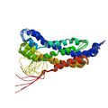
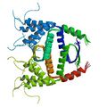
Reckel, S., Gehin, C., Tardivon, D., Harduin, D., Georgeon, S., Kükenshöner, T., Löhr, F., Koide, A., Buchner, L., Panjkovich, A., Reynaud, A., Pinho, S., Gerig, B., Svergun, D., Pojer, F., Güntert, P., Dötsch, V., Koide, S., Gavin, A.-C. & Hantschel, O. Structural and functional dissection of the DH and PH domains of oncogenic Bcr-Abl tyrosine kinase. Nat. Commun. 8, 2101 (2017)
PDB 5N6R NMR restraints BMRB 34101 (DH NMR structure) |
|
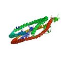
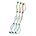
Bibow, S., Polyhach, Y., Eichmann, C., Chi, C. N., Kowal, J., Albiez, S., McLeod, R. A., Stahlberg, H., Jeschke, G., Güntert, P. & Riek, R. Solution structure of discoidal high-density lipoprotein particles with a shortened apolipoprotein A-I. Nat. Struct. Mol. Biol. 24, 187-193 (2017)
PDB 2N5E NMR restraints BMRB 25710 |
|
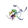
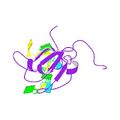
Kuwasako, K., Nameki, N., Tsuda, K., Takahashi, M., Sato, A., Tochio, N., Inoue, M., Terada, T., Kigawa, T., Kobayashi, N., Shirouzu, M., Ito, T., Sakamoto, T., Wakamatsu, K., Güntert, P., Takahashi, S., Yokoyama, S. & Muto, Y. Solution structure of the first RNA recognition motif domain of human spliceosomal protein SF3b49 and its mode of interaction with a SF3b145 fragment. Protein Sci. 26, 280-291 (2017)
PDB 5GVQ NMR restraints BMRB 36018 |
|
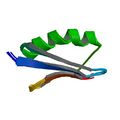
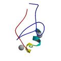
Ikeya, T., Hanashima, T., Hosoya, S., Shimazaki, M., Ikeda, S., Mishima, M., Güntert, P. & Ito, Y. Improved in-cell structure determination of proteins at near-physiological concentration. Sci Rep. 6, 38312 (2016)
PDB 2N9L NMR restraints BMRB 25910 |
|
Gebel, J., Luh, L. M., Coutandin, D., Osterburg, C., Löhr, F., Schäfer, B., Frombach, A., Sumyk, M., Buchner, L., Krojer, T., Salah, E., Mathea, S., Güntert, P., Knapp, S. & Dötsch, V. Mechanism of TAp73 inhibition by ΔNp63 and structural basis of p63/p73 hetero-tetramerization. Cell Death Diff. 23, 1930-1940 (2016)
PDB 2NB1 NMR restraints BMRB 25958 |
|
Wälti, M. A., Ravotti, F., Arai, H., Glabe, C., Wall, J., Böckmann, A., Güntert, P., Meier, B. H. & Riek, R. Atomic resolution structure of a disease-relevant Aβ(1–42) amyloid fibril. Proc. Natl. Acad. Sci. USA
PDB 2NAO NMR restraints BMRB 26692 |

|
von Delbrück, M., Kniss, A., Rogov, V. V., Pluska, L., Bagola, K., Löhr, F., Güntert, P., Sommer, T. & Dötsch, V. The CUE domain of Cue1 aligns growing ubiquitin chains with Ubc7 for rapid elongation. Mol. Cell 62, 918-928 (2016)
PDB 2MYX NMR restraints BMRB 25461 |
|
Huang, S. Y., Chang, C. F., Fan, P. J., Naik, M. T., Güntert, P., Shih, H. M. & Huang, T. H. The RING domain of human promyelocytic leukemia protein (PML). J. Biomol. NMR 61, 173-180 (2015)
PDB 2MWX NMR restraints BMRB 25376 |

|
Schütz, A. K., Vagt, T., Huber, M., Ovchinnikova, O. Y., Cadalbert, R., Wall, J., Güntert, P., Böckmann, A., Glockshuber, R., and Meier, B. H. Atomic-resolution three-dimensional structure of amyloid β fibrils bearing the Osaka mutation. Angew. Chem. Int. Ed. 54, 331–335 (2015)
PDB 2MVX NMR restraints BMRB 25289 |

|
Tsuda, K., Kuwasako, K., Nagata, T., Takahashi, M., Kigawa, T., Kobayashi, N., Güntert, P., Shirouzu, M., Yokoyama, S. & Muto, Y. Novel RNA recognition motif domain in cytoplasmic polyadenylation element binding protein 3. Proteins 82, 2879–2886 (2014)
PDB 2RUG NMR restraints BMRB 11563 |
|
Kuwasako, K., Takahashi, M., Unzai, S., Tsuda, K., Yoshikawa, S., He, F., Kobayashi, N., Güntert, P., Shirouzu, M., Ito, T., Tanaka, A., Yokoyama, S., Hagiwara, M., Kuroyanagi, H. & Muto, Y. RBFOX and SUP-12 sandwich a guanine base to cooperatively regulate tissue-specific splicing. Nat. Struct. Mol. Biol. 21, 778–786 (2014)
PDB 2RU3 NMR restraints BMRB 11517 (SUP-12–RNA6 complex) |
|
Uggerhøj, L. E., Munk, J. K., Hansen, P. R., Güntert, P. & Wimmer, R. Structural features of peptoid-peptide hybrids in lipid-water interfaces. FEBS Lett. 588, 3291–3297 (2014)
PDB 2MMJ NMR restraints BMRB 19856 (M-Nleu11) |
|
Watson, R. P., Christen, M. T., Bumbak, F., Ewald, C., Reichen, C. Mihajlovic, M., Schmidt, E., Güntert, P., Caflisch, A., Plückthun, A., Zerbe, O. Spontaneous self assembly of fragments of engineered Armadillo repeat proteins into a folded structure. Structure 22, 985–995 (2014)
PDB 2RU5 NMR restraints BMRB 11548 (uncomplexed MA) |
|
Tufar, P., Rahighi, S., Kraas, F. I., Kirchner, D. K., Löhr, F., Henrich, E., Köpke, J., Dikic, I., Güntert, P., Marahiel, M. A. & Dötsch, V. Crystal structure of a PCP/Sfp complex reveals the structural basis for carrier protein posttranslational modification. Chem. Biol. 21, 552–562 (2014)
PDB 2MD9 NMR restraints BMRB 19479 |
|
Kogure, H., Handa, Y., Nagata, M., Kanai, N., Güntert, P., Kubota, K. & Nameki, N. Identification of residues required for stalled-ribosome rescue in the codon-independent release factor YaeJ. Nucl. Acids Res. 42, 3152-3163 (2014)
PDB 2RTX NMR restraints BMRB 11534 |
|
Luh, L. M., Hänsel, R., Löhr, F., Kirchner, D. K., Krauskopf, K., Pitzius, S., Schäfer, B., Tufar, P., Corbeski, I., Güntert, P. & Dötsch, V. Molecular crowding drives active Pin1 into nonspecific complexes with endogenous proteins prior to substrate recognition. J. Am. Chem. Soc. 135, 13796−13803 (2013)
PDB 2M8I NMR restraints BMRB 19258 (wildtype
WW domain) |
|
Rogov, V. V., Suzuki, H., Fiskin, E., Wild, P., Kniss, A., Rozenknop, A., Kato, R., Kawasaki, M., McEwan, D. G., Löhr, F., Güntert, P., Dikic, I., Wakatsuki, S. & Dötsch, V. Structural basis for phosphorylation-triggered autophagic clearance of Salmonella. Biochem. J. 454, 459–466 (2013)
PDB 2LUE NMR restraints BMRB 18518 (NMR structure of LC3B OPTN-LIR Ptot complex) |
|
Lin, Y. J., Ikeya, T., Güntert, P. & Chang, L. S. NMR solution structure of a chymotrypsin inhibitor from the Taiwan cobra. Molecules 18, 8906-8918 (2013)
PDB 2M99 NMR restraints BMRB 19287 |
|
He, F., Tsuda, K., Takahashi, M., Kuwasako, K., Terada, T., Shirouzu, M., Watanabe, S., Kigawa, T., Kobayashi, N., Güntert, P., Yokoyama, S. & Muto, Y. Structural insight into the interaction of ADP-ribose with the PARP WWE domains. FEBS Lett. 586, 3858–3864 (2012)
PDB 2DK6 NMR restraints BMRB 11500 (WWE domain from PARP11) |
|
Christ, N. A., Bochmann, S., Gottstein, D., Duchardt-Ferner, E., Hellmich, U. A., Düsterhus, S., Kötter, P., Güntert, P., Entian, K.-D. & Wöhnert, J. The first structure of a lantibiotic immunity protein, SpaI from Bacillus subtilis, reveals a novel fold. J. Biol. Chem. 287, 35286-35298 (2012)
PDB 2LVL NMR restraints BMRB 17534 |
|
Vögeli, B., Kazemi, S., Güntert, P. & Riek, R. Spatial elucidation of motion in proteins by ensemble-based structure calculation using exact NOEs. Nature Struct. Mol. Biol. 19, 1053–1057 (2012)
PDB 2LUM NMR restraints BMRB 18531 |
|
Kogure, H., Hikawa, Y., Hagihara, M., Tochio, N., Koshiba, S., Inoue, Y., Güntert, P., Kigawa, T., Yokoyama, S. & Nameki, N. Solution structure and siRNA-mediated knockdown analysis of the mitochondrial disease-related protein C12orf65. Proteins 80, 2629–2642 (2012)
PDB 2RSM NMR restraints BMRB 11491 |
|
Nagata, T., Tsuda, K., Shirouzu, M., Kigawa, T., Kobayashi, N., Güntert, P., Yokoyama, S. & Muto, Y. Solution structures of the double-stranded RNA-binding domains from RNA-helicase A. Proteins 80, 1699–1706 (2012)
PDB 2RS6 NMR restraints BMRB 11456 (dsRBD1) |
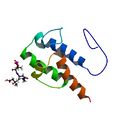
|
Busche, A., Gottstein, D., Hein, C., Ripin, N., Pader, I., Tufar, P., Eisman, E. B., Gu, L. Walsh, C. T., Sherman, D. H., Löhr, F., Güntert, P. & Dötsch, V. Characterization of the interaction between an ACP domain and a halogenase in the curacin A polyketide synthetase. ACS Chem. Biol. 7, 378–386 (2012)
PDB 2LIU NMR restraints BMRB 17906 (holo-ACPI) |
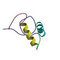
|
He, F., Inoue, M., Kigawa, T., Takahashi, M., Kuwasako, K., Tsuda, K., Kobayashi, N., Terada, T., Shirouzu, M., Tanaka, A., Sugano, S., Güntert, P., Yokoyama, S. & Muto, Y. Solution structure of the Splicing Factor Motif (SFM) of the human Prp18 protein. Proteins 80, 968–974 (2012)
PDB 2DK4 NMR restraints BMRB 11355 |
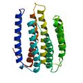
|
Reckel, S., Gottstein, D., Stehle, J., Löhr, F., Verhoefen, M. K., Takeda, M., Silvers, R., Kainosho, M., Glaubitz, C., Wachtveitl, J., Bernhard, F., Schwalbe, H., Güntert, P. & Dötsch, V. Solution NMR structure of proteorhodopsin. Angew. Chem. Int. Ed. 50, 11942–11946 (2011)
PDB 2L6X NMR restraints BMRB 17327 |
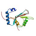
|
Rozenknop, A., Rogov, V. V., Löhr, F., Güntert, P., Dikic, I., & Dötsch, V. Characterization of the interaction of GABARAPL-1 with the LIR motif of NBR1. J. Mol. Biol. 410, 477–487 (2011)
PDB 2L8J NMR restraints BMRB 17412 |
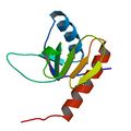
|
Schmöe, K., Rogov, V. V., Rogova, N. Y., Löhr, F., Güntert, P., Bernhard, F. & Dötsch, V. Structural insights into Rcs phosphotransfer: The newly identified RcsD-ABL domain enhances interaction with the response regulator RcsB. Structure 19, 577–587 (2011)
PDB 2KX7 NMR restraints |
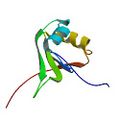
|
Tsuda, K., Someya, T., Kuwasako, K., Takahashi, M., Fahu, H., Inoue, M., Harada, T., Watanabe, S., Terada, T., Kobayashi, N., Shirouzu, M., Kigawa, T., Tanaka, A., Sugano, S., Güntert, P., Yokoyama, S. & Muto, Y. Structural basis for the dual RNA-recognition modes of human Tra2-β RRM. Nucl. Acids Res. 39, 1538-1553 (2011)
PDB 2RRB NMR restraints |
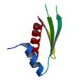
|
Yamashita, S., Nagata, T., Kawazoe, M., Takemoto, C., Kigawa, T, Güntert, P., Kobayashi, N., Terada, T., Shirouzu, M., Wakiyama, M., Muto, Y. & Yokoyama, S. Structures of the first and second double-stranded RNA-binding domains of human TAR RNA-binding protein. Protein Sci. 20, 118-130 (2011) |
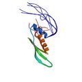
|
Yamashita, S., Nagata, T., Kawazoe, M., Takemoto, C., Kigawa, T, Güntert, P., Kobayashi, N., Terada, T., Shirouzu, M., Wakiyama, M., Muto, Y. & Yokoyama, S. Structures of the first and second double-stranded RNA-binding domains of human TAR RNA-binding protein. Protein Sci. 20, 118-130 (2011)
PDB 2CPN NMR restraints |
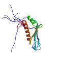
|
Handa, Y., Hikawa, Y., Tochio, N., Kogure, H., Inoue, M., Koshiba, S., Güntert, P., Inoue, Y., Kigawa, T., Yokoyama, S. & Nameki, N. Solution structure of the mitochondrial protein ICT1 that is essential for cell vitality. J. Mol. Biol. 404, 260-273 (2010)
PDB 1J26 |
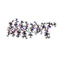
|
Hamada, T., Matsunaga, S.,Fujiwara, M., Fujita, K., Hirota, H., Schmucki, R., Güntert, P. & Fusetani, N. Solution structure of polytheonamide B, a highly cytotoxic nonribosomal polypeptide from marine sponge. J. Am. Chem. Soc. 132, 12941–12945 (2010)
PDB 2RQO NMR restraints |
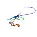
|
He, F., Umehara, T., Saito, K., Harada, T., Watanabe, S., Yobuki, T., Kigawa, T., Takahashi, M., Kuwasako, K., Tsuda, K., Matsuda, T., Aoki, M., Seki, E., Kobayashi, N., Güntert, P., Yokoyama, S. & Muto, Y. Structural insight into the zinc finger CW domain as a histone modification reader. Structure 18, 1127–1139 (2010)
PDB 2E61 NMR restraints |
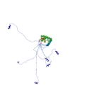
|
Sobhanifar, S., Schneider, B., Löhr, F., Gottstein, D., Ikeya, T., Filipek, S., Güntert, P., Bernhard, F. & Dötsch, V. Structural investigation of the C-terminal catalytic fragment of presenilin-1. Proc. Natl. Acad. Sci. USA 107, 9644–9649 (2010)
PDB 2KR6 NMR restraints |
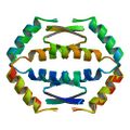
|
Coutandin, D., Löhr, F., Niesen, F. H., Ikeya, T., Weber, T. A., Schäfer, B., Bullock, A. N., Yang, A., Güntert, P. , Knapp, S., McKeon, F., Der Ou, H. & Dötsch, V. Conformational stability and activity of p73 require a second helix in the tetramerization domain. Cell Death Diff. 16, 1582–1589 (2009)
PDB 2KBY NMR restraints |
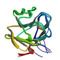
|
He, F., Saito, K., Kobayashi, N., Harada, T., Watanabe, S., Kigawa, T, Güntert, P., Unzai, S., Muto, Y. & Yokoyama, S. Structural and functional characterization of the NHR1 domain of the Drosophila Neuralized E3 ligase in the Notch signaling pathway. J. Mol. Biol. 393, 478-495 (2009)
PDB 2YUE NMR restraints |
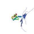
|
Khayrutdinov, B. I., Bae, W. J., Yun, Y. M., Lee, J. H., Tsuyama, T., Kim, J. J., Hwang, E., Ryu, K. S., Cheong, H. K., Cheong, C., Ko, J. S., Enomoto, T., Karplus, P. A., Güntert, P., Tada, S., Jeon, Y. H., Cho, Y. Structure of the Cdt1 C-terminal domain: Conservation of the winged helix fold in replication licensing factors. Protein Sci. 18, 2252-2264 (2009)
PDB 2KLO NMR restraints |
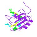
|
Tsuda, K., Kuwasako, K., Takahashi, M., Someya, T., Inoue, M., Terada, T., Kobayashi, N., Shirouzu, M., Kigawa, T., Tanaka, A., Sugano, S., Güntert, P., Muto, Y. & Yokoyama, S. Structural basis for the sequence specific RNA-recognition mechanism of human CUG-BP1 RRM3. Nucl. Acids Res. 37, 5151–5166 (2009)
PDB 2RQC NMR restraints |
|
He, F., Dang, W., Saito, K., Watanabe, S., Kobayashi, N., Güntert, P., Kigawa, T, Tanaka, A., Muto, Y. & Yokoyama, S. Solution structure of the cysteine-rich domain in Fn14, a member of the tumor necrosis factor receptor superfamily. Protein Sci. 18, 650–656 (2009)
PDB 2RPJ NMR restraints |
|
Peroza, E. A., Schmucki, R., Güntert, P., Freisinger, E. & Zerbe O. The βE-domain of Wheat Ec-1 metallothionein: A metal-binding domain with a distinctive structure. J. Mol. Biol. 387, 207–218 (2009)
PDB 2KAK NMR restraints BMRB 16025 |
|
Sakakibara, D., Sasaki, A., Ikeya, T., Hamatsu, J., Hanashima, T., Mishima, M., Yoshimasu, M., Hayashi, N., Mikawa, T., Wälchli, M., Smith, B. O., Shirakawa, M., Güntert, P. & Ito, Y. Protein structure determination in living cells by in-cell NMR spectroscopy. Nature 458, 102-105 (2009)
PDB 2ROG NMR restraints BMRB 11037 (in-cell) |
|
He, F., Dang, W., Abe, C., Tsuda, K., Inoue, M., Watanabe, S., Kobayashi, N., Kigawa, T, Matsuda, T., Yabuki, T., Aoki, M., Seki, E., Harada, T., Tomabechi, Y., Terada, T., Shirouzu, M., Tanaka, A., Güntert, P., Muto, Y. & Yokoyama, S. Solution structure of the RNA binding domain in the human muscleblind-like protein 2. Protein Sci. 18, 80-91 (2009)
PDB 2E5S NMR restraints |

|
Ohnishi, S. Pääkkönen, K., Koshiba, S., Tochio, N., Sato, M., Kobayashi, N., Harada, T., Watanabe, S., Muto, Y., Güntert, P., Tanaka, A., Kigawa, T. & Yokoyama, S. Solution structure of the GUCT domain from human RNA helicase II/Guβ reveals the RRM fold, but implausible RNA interactions. Proteins 74, 133–144 (2009)
PDB 2E29 |
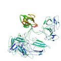
|
Takeda, M., Sugimori, N., Torizawa, T., Terauchi, T., Ono, A. M., Yagi, H., Yamaguchi, Y., Kato, K., Ikeya, T., Jee, J., Güntert, P., Aceti, D. J., Markley, J. L. & Kainosho, M. Structure of the putative 32 kDa myrosinase binding protein from Arabidopsis (At3g16450.1) as determined by the SAIL-NMR method. FEBS J. 275, 5873–5884 (2008)
PDB 2JZ4 NMR restraints BMRB 15607 |
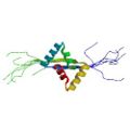
|
Yoshida, H., Furuya, N., Lin, Y. J., Güntert, P., Komano, T. & Kainosho, M. Structural basis of the role of the NikA ribbon-helix-helix domain in initiating bacterial conjugation. J. Mol. Biol. 384, 690–701 (2008) |
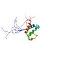
|
Lin, Y. J., Umehara, T., Inoue, M., Saito, K., Kigawa, T., Jang, M. K., Ozato, K., Yokoyama, S., Padmanabhan, B., Güntert, P. Solution structure of the extraterminal domain of the bromodomain-containing protein BRD4. Protein Sci. 17, 2174–2179 (2008) |
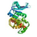
|
Koglin, A., Löhr, F., Bernhard, F., Rogov, V. R., Frueh, D. P., Strieter, E. R., Mofid, M. R., Güntert, P., Wagner, G., Walsh, C. T., Marahiel, M. A. & Dötsch, V. Structural basis for the selectivity of the external thioesterase of the surfactin-synthetase. Nature 454, 907–911 (2008)
PDB 2RON NMR restraints (SrfTEII) |
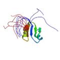
|
Nagata, T., Suzuki, S., Endo, R., Shirouzu, M., Terada, T., Inoue, M., Kigawa, T, Güntert, P., Hayashizaki, Y., Muto, Y. & Yokoyama, S. The RRM domain of poly(A)-specific ribonuclease (PARN) has a non-canonical binding site for mRNA cap analog recognition. Nucl. Acids Res. 36, 4754–4767 (2008) |
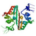
|
Takeda, M., Chang, C. K., Ikeya, T., Güntert, P., Chang, Y. H., Hsu, Y. L., Huang, T. H. & Kainosho, M. Solution structure of the C-terminal dimerization domain of SARS coronavirus nucleocapsid protein determined by the SAIL-NMR method. J. Mol. Biol. 380, 608–622 (2008) |
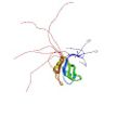
|
Kuwasako, K., Takahashi, M., Tochio, N., Abe, C., Tsuda, K., Inoue, M., Terada, T., Shirouzu, M., Kobayashi, N., Kigawa, T., Taguchi, S., Tanaka, A., Hayashizaki, Y., Güntert, P., Muto, Y. & Yokoyama, S. Solution structure of the second RNA recognition motif (RRM) domain of murine T cell intracellular antigen-1 (TIA-1) and its RNA recognition mode. Biochemistry 47, 6437–6450 (2008)
PDB 2RNE |
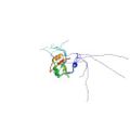
|
Kuwasako, K., Dohmae, N., Inoue, M., Shirouzu, M., Taguchi, S., Güntert, P., Séraphin, B., Muto, Y. & Yokoyama, S. Complex assembly mechanism and an RNA-binding mode of the human p14-SF3b155 spliceosomal protein complex identified by NMR solution structure and functional analyses. Proteins 71, 1617–1636 (2008)
PDB 2FHO NMR restraints |
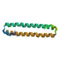
|
Hwang, E., Ryu, K. S., Pääkkönen, K., Güntert, P., Cheong, H. K., Lim, D. S., Lee, J. O., Jeon, Y. H. & Cheong, C. Structural insight into dimeric interaction of the SARAH domains from Mst1 and RASSF family proteins in the apoptosis pathway. Proc. Natl. Acad. Sci. USA. 104, 9236–9241 (2007)
PDB 2JO8 NMR restraints |
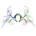
|
Ohnishi, S. Güntert, P., Koshiba, S., Tomizawa, T., Akasaka, R., Tochio, N., Sato, M., Inoue, M., Harada, T., Watanabe, S., Tanaka, T., Shirouzu, M., Kigawa, T. & Yokoyama, S. Solution structure of an atypical WW domain in a novel β-clam-like dimeric form. FEBS Lett. 581, 462–468 (2007)
PDB 2DWV |

|
Kuwasako, K., He, F., Inoue, M., Tanaka, A., Sugano, S., Güntert, P., Muto, Y. & Yokoyama, S. Solution structures of the SURP domains and the subunit-assembly mechanism within the splicing factor SF3a complex in 17S U2 snRNP. Structure 14, 1677–1689 (2006) |

|
ENTH-VHS domain At3g16270(9-135) from Arabidopsis thaliana.
López-Méndez, B. & Güntert, P. Automated protein structure determination from NMR spectra. J. Am. Chem. Soc. 128, 13112–13122 (2006) PDB 2DCP NMR restraints Note: Automated structure determination with FLYA. Structure does not supersede the original deposition 1VDY. |

|
Rhodanese homology domain At3g16270(175-295) from Arabidopsis thaliana.
López-Méndez, B. & Güntert, P. Automated protein structure determination from NMR spectra. J. Am. Chem. Soc. 128, 13112–13122 (2006) PDB 2DCQ NMR restraints Note: Automated structure determination with FLYA. Structure does not supersede the original deposition 1VEE. |

|
Src homology domain 2 from the human feline sarcoma oncogene Fes.
López-Méndez, B. & Güntert, P. Automated protein structure determination from NMR spectra. J. Am. Chem. Soc. 128, 13112–13122 (2006) PDB 2DCR NMR restraintsbr>Note: Automated structure determination with FLYA. Structure does not supersede the original deposition 1WQU. |

|
Jurt, S., Aemissegger, A., Güntert, P., Zerbe, O. & Hilvert, D. A photoswitchable miniprotein based on the sequence of avian pancreatic polypeptide. Angew. Chem. Int. Ed. 45, 6297-6300 (2006)
PDB 2H4B NMR restraints (cis-1-PP) |

|
Hamada, T., Ito, Y., Abe, T., Hayashi, F., Güntert, P., Inoue, M., Kigawa, T., Terada, T., Shirouzu, M., Yoshida, M., Tanaka, A., Sugano, S., Yokoyama, S. & Hirota, H. Solution structure of the antifreeze-like domain of human sialic acid synthase. Protein Sci. 15, 1010–1016 (2006)
PDB 1WVO NMR restraints |

|
Aachmann, F. L., Svanem, B. I. G., Güntert, P., Petersen, S. B., Valla, S. & Wimmer, R. NMR structure of the R-module - A parallel β-roll subunit from an Azotobacter vinelandii mannuronan C-5 epimerase. J. Biol. Chem. 281, 7350–7356 (2006)
PDB 2AGM NMR restraints |

|
Stereo-array isotope labeled (SAIL) maltodextrin-binding protein MBP.
Kainosho, M., Torizawa, T., Iwashita, Y., Terauchi, T., Ono, A. M. & Güntert, P. Optimal isotope labeling for NMR protein structure determinations. Nature 440, 52–57 (2006) |

|
Stereo-array isotope labeled (SAIL) calmodulin.
Kainosho, M., Torizawa, T., Iwashita, Y., Terauchi, T., Ono, A. M. & Güntert, P. Optimal isotope labeling for NMR protein structure determinations. Nature 440, 52–57 (2006) |

|
Li, H., Inoue, M., Yabuki, T., Aoki, M., Seki, E., Matsuda, T., Nunokawa, E., Motoda, Y., Kobayashi, A., Terada, T., Shirouzu, M., Koshiba, S., Lin, Y. J., Güntert, P., Suzuki, H., Hayashizaki, Y., Kigawa, T. & Yokoyama, S. Solution structure of the mouse enhancer of rudimentary protein reveals a novel fold. J. Biomol. NMR 32, 329–334 (2005)
PDB 1WWQ |

|
Scott, A., Pantoja-Uceda, D., Koshiba, S., Inoue, M., Kigawa, T., Terada, T., Shirouzu, M., Tanaka, A., Sugano, S., Yokoyama, S. & Güntert, P. Solution structure of the Src homology 2 domain from the human feline sarcoma oncogene Fes. J. Biomol. NMR. 31, 357–361 (2005) |

|
Nameki, N., Tochio, N., Koshiba, S., Inoue, M., Yabuki, T., Aoki, M., Seki, E., Matsuda, T., Fujikura, Y., Saito, M., Ikari, M., Watanabe, M., Terada, T., Shirouzu, M., Yoshida, M., Hirota, H., Tanaka, A., Hayashizaki, Y., Güntert, P., Kigawa, T. & Yokoyama, S. Solution structure of the PWWP domain of the hepatoma-derived growth factor family. Protein Sci. 14, 756–764 (2005)
PDB 1N27 |

|
Calzolai, L., Lysek, D. A., Pérez, D. R., Güntert, P. & Wüthrich, K. Prion protein NMR structures of chicken, turtle and frog. Proc. Natl. Acad. Sci. USA 102, 651–655 (2005)
PDB 1U3M NMR restraints BMRB 6269 (chicken PrP(121-225)) |

|
Lysek, D. A., Schorn, C., Nivon, L. G., Esteve-Moya, V., Christen, B., Calzolai, L., von Schroetter, C., Fiorito, F., Herrmann, T., Güntert, P. & Wüthrich, K. Prion protein NMR structures of cat, dog, pig and sheep. Proc. Natl. Acad. Sci. USA 102, 640–645 (2005)
PDB 1XYJ NMR restraints BMRB 6377 (cat PrP(121-231)) |

|
Pääkkönen, K., Tossavainen, H., Permi, P., Rakkolainen, H., Rauvala, H., Raulo, E., Kilpeläinen, I. & Güntert, P. Solution structures of the first and fourth TSR domains of F-spondin. Proteins 64, 665–672 (2006)
PDB 1SZL NMR restraints BMRB 6175 (TSR1) |

|
ENTH-VHS domain At3g16270 from Arabidopsis thaliana. |

|
Iwai, H., Forrer, P., Plückthun, A. & Güntert, P. NMR solution structure of the monomeric form of the bacteriophage λ capsid stabilizing protein gpD. J. Biomol. NMR. 31, 351–356 (2005) |

|
Pantoja-Uceda, D., López-Méndez, B., Koshiba, S., Inoue, M., Kigawa, T., Terada, T., Shirouzu, M., Tanaka, A., Seki, M., Shinozaki, K., Yokoyama, S. & Güntert, P. Solution structure of the rhodanese homology domain At4g01050(175–295) from Arabidopsis thaliana. Protein Sci. 14, 224–230 (2005) |

|
Nameki, N., Yoneyama, M., Koshiba, S., Tochio, N., Inoue, M., Seki, E., Matsuda, T., Tomo, Y., Harada, T., Saito, K., Kobayashi, N., Yabuki, T., Aoki, M., Nunokawa, E., Matsuda, N., Sakagami, N., Terada, T., Shirouzu, M., Yoshida, M., Hirota, H., Osanai, T., Tanaka, A., Arakawa, T., Carninci, P., Kawai, J., Hayashizaki, Y., Kinoshita, K., Güntert, P., Kigawa, T. & Yokoyama, S. Solution structure of the RWD domain of the mouse GCN2 protein. Protein Sci. 13, 2089–2100 (2004) |

|
Fernández, C., Hilty, C., Wider, G., Güntert, P. & Wüthrich, K. NMR structure of the integral membrane protein OmpX. J. Mol. Biol. 336, 1211–1221 (2004) |

|
FHA domain of mouse afadin 6.
PDB 1WLN |

|
Vanwetswinkel, S., Kriek, J., Andersen, G. R., Güntert, P., Dijk, P., Canters, G. W. & Siegal, G. Solution structure of the 162 residue C-terminal domain of human elongation factor 1Bγ. J. Biol. Chem. 278, 43443–43451 (2003)
PDB 1PBU NMR restraints BMRB 5628 |

|
Hilge, M., Siegal, G., Vuister, G. W., Güntert, P., Gloor, S. M. & Abrahams, J. P. ATP-induced conformational changes of the nucleotide-binding domain of Na,K-ATPase. Nat. Struct. Biol. 10, 468–474 (2003)
PDB 1MO7 BMRB 5577 (free form) |

|
Lührs, T., Riek, R., Güntert, P. & Wüthrich, K. NMR structure of the human doppel protein. J. Mol. Biol. 326, 1549–1557 (2003)
PDB 1LG4 NMR restraints BMRB 5145 |

|
Zahn, R., Güntert, P. & Wüthrich, K. NMR structure of a variant human prion protein with two disulfide bonds. J. Mol. Biol. 326, 225–234 (2003)
PDB 1H0L NMR restraints BMRB 5378 |

|
Enggist, E., Thöny-Meyer, L., Güntert, P. & Pervushin, K. NMR structure of the heme chaperone CcmE reveals a new functional motif. Structure 10, 1551–1557 (2002)
PDB 1SR3 NMR restraints |

|
Lee, D., Damberger, F. D., Peng, G., Horst, R., Güntert, P., Nikonova, L., Leal, W. S. & Wüthrich, K. NMR structure of the unliganded Bombyx mori pheromone-binding protein at physiological pH. FEBS Lett. 531, 314–318 (2002)
PDB 1LS8 |

|
Ellgaard, L., Bettendorff, P., Braun, D., Herrmann, T., Fiorito, F., Jelesarov, I., Güntert, P., Helenius, A. & Wüthrich, K. NMR Structures of 36 and 73-residue fragments of the calreticulin P-domain. J. Mol. Biol. 322, 773–784 (2002)
PDB 1K9C NMR restraints BMRB 5204 (residues 189-261) |

|
Miura, T., Klaus, W., Ross, A., Güntert, P., Senn, H. The NMR structure of the class I human ubiquitin-conjugating enzyme 2b. J. Biomol. NMR 22, 89–92 (2002)
PDB 1JAS NMR restraints BMRB 5038 |

|
Horst, R., Damberger, F., Luginbühl, P., Güntert, P., Peng, G., Nikonova, L., Leal, W. S. & Wüthrich, K. NMR structure reveals intramolecular regulation mechanism for pheromone binding and release. Proc. Natl. Acad. Sci. USA 98, 14374–14379 (2001)
PDB 1GM0 NMR restraints BMRB 4849 |

|
Ellgaard, L., Riek, R., Herrmann, T., Güntert, P., Braun, D., Helenius, A. & Wüthrich, K. NMR structure of the calreticulin P-domain. Proc. Natl. Acad. Sci. USA 98, 3133–3138 (2001) |

|
Calzolai, L., Lysek, D. A., Güntert, P., von Schroetter, C., Riek, R., Zahn, R. & Wüthrich, K. NMR structures of three single-residue variants of the human prion protein. Proc. Natl. Acad. Sci. USA 97, 8340–8345 (2000)
PDB 1E1G NMR restraints BMRB 4736 (variant M166V) |

|
Riek, R., Prêcheur, B., Wang, Y., Wider, G., Güntert, P., Liu, A., Kägi, J. H. R. & Wüthrich, K. NMR structure of the sea urchin (Strongylocentrotus purpuratus) metallothionein MTA. J. Mol. Biol. 291, 417–428 (1999)
PDB 1QJK NMR restraints (α domain) |

|
Pellecchia, M., Güntert, P., Glockshuber, R. & Wüthrich, K. The NMR solution structure of the periplasmic chaperone FimC. Nature Struct. Biol. 5, 885–890 (1998)
PDB 1BF8 NMR restraints BMRB 4070 |

|
Ottiger, M., Zerbe, O., Güntert, P. & Wüthrich, K. The NMR solution conformation of unligated human Cyclophilin A. J. Mol. Biol. 272, 64–81 (1997)
PDB 1OCA NMR restraints |

|
Antuch, W., Güntert, P. & Wüthrich, K. Ancestral βγ-crystallin precursor structure in a yeast killer toxin. Nature Struct. Biol. 3, 662–665 (1996)
PDB 1WKT NMR restraints BMRB 5255 |

|
Antuch, W., Güntert, P., Billeter, M., Hawthorne, T., Grossenbacher, H. & Wüthrich, K. NMR solution structure of the recombinant tick anticoagulant protein (rTAP), a factor Xa inhibitor from the tick Ornithodoros moubata. FEBS Lett. 352, 251–257 (1994)
PDB 1TAP NMR restraints |

|
Berndt, K. D., Güntert, P. & Wüthrich, K. The NMR solution structure of dendrotoxin K from the venom of Dendroaspis polylepis polylepis. J. Mol. Biol. 234, 735–750 (1993)
PDB 1DTK NMR restraints |

|
Szyperski, T., Güntert, P., Stone, S. R. & Wüthrich, K. NMR solution structure of hirudin(1–51) and comparison with corresponding three-dimensional structures determined using the complete 65-residue hirudin polypeptide chain. J. Mol. Biol. 228, 1193–1205 (1992)
PDB 1HIC NMR restraintsBMRB 1128 |

|
Berndt, K. D., Güntert, P., Orbons, L. P. M. & Wüthrich, K. Determination of a high-quality NMR solution structure of the bovine pancreatic trypsin inhibitor (BPTI) and comparison with three crystal structures. J. Mol. Biol. 227, 757–775 (1992)
PDB 1PIT NMR restraints |

|
Güntert, P., Qian, Y. Q., Otting, G., Müller, M., Gehring, W. J. & Wüthrich K. Structure determination of the Antp(C39S) homeodomain from nuclear magnetic resonance data in solution using a novel strategy for the structure calculation with the programs DIANA, CALIBA, HABAS and GLOMSA. J. Mol. Biol. 217, 531–540 (1991)
PDB 2HOA |